Copyright
©The Author(s) 2023.
World J Gastroenterol. Sep 14, 2023; 29(34): 5082-5090
Published online Sep 14, 2023. doi: 10.3748/wjg.v29.i34.5082
Published online Sep 14, 2023. doi: 10.3748/wjg.v29.i34.5082
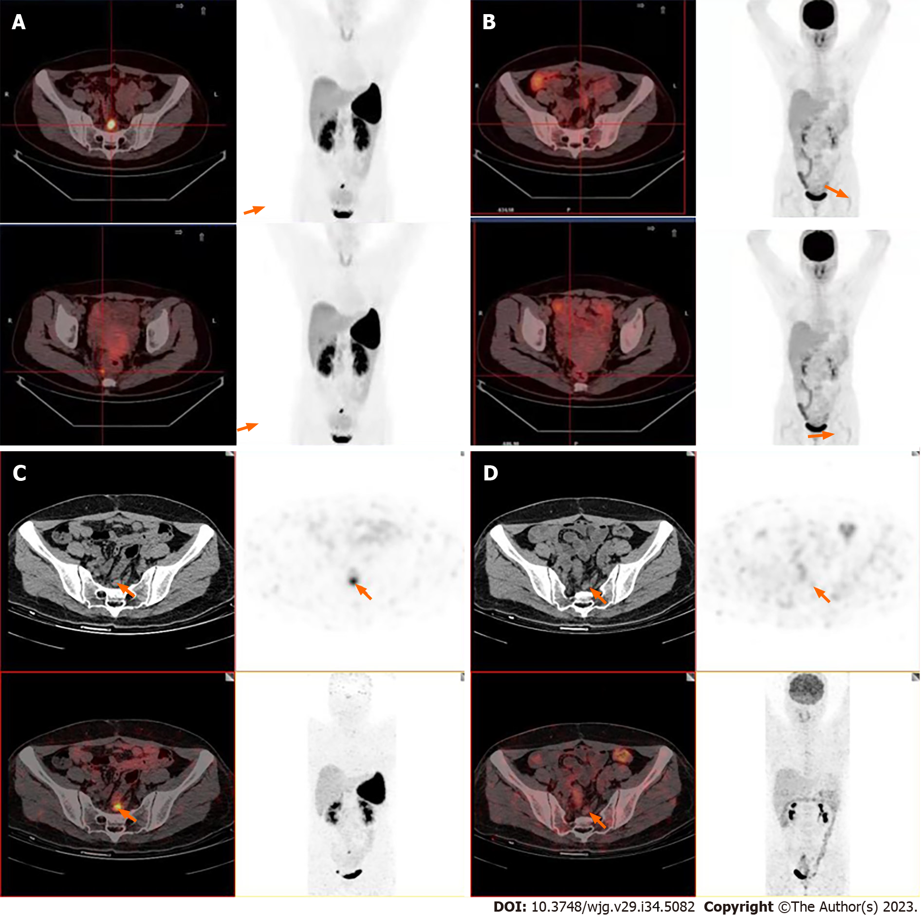
Figure 1 Images.
A and B: 18F-ALF-NOTATATE positron emission tomography-computed tomography (PET/CT) images and corresponding 18F-fluorodeoxyglucose (18F-FDG) PET/CT images in a patient with rectal neuroendocrine tumors in our hospital; C and D: 68Ga-DOTANOC PET/CT and 18F-FDG PET/CT images in the First Affiliated Hospital of Sun Yat-sen University.
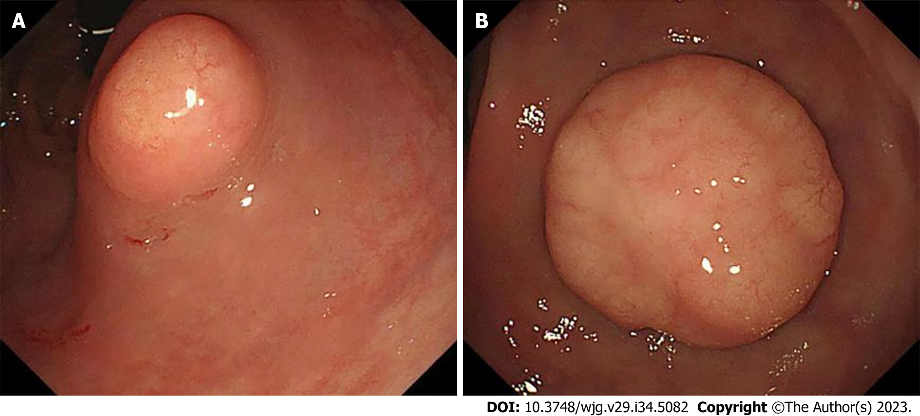
Figure 2 Two lesions.
A: The one lesion at the lower rectum under colonoscopy; B: The second lesion at the lower rectum under colonoscopy.
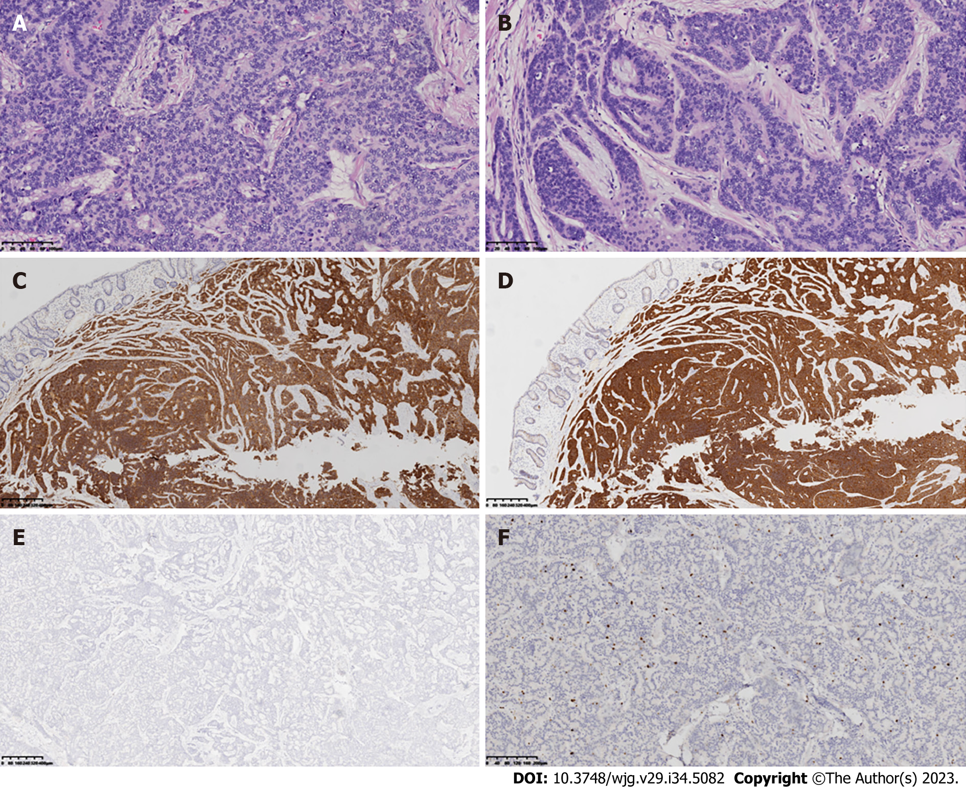
Figure 3 Hematoxylin-eosin staining and immunohistochemical staining.
A and B: Hematoxylin-eosin staining shows the pathological features of rectal neuroendocrine tumors in the larger and smaller tumor tissue specimens (20 ×); C-E: Immunohistochemical staining of neuroendocrine markers CD56, Syn, and CgA in representative tumor tissue selected by the physician (4 ×); F: Ki-67 expression in the neuroendocrine tumor at the rectum by immunohistochemistry (10 ×). Brown nuclear stain highlights Ki-67-positive tumor cells.
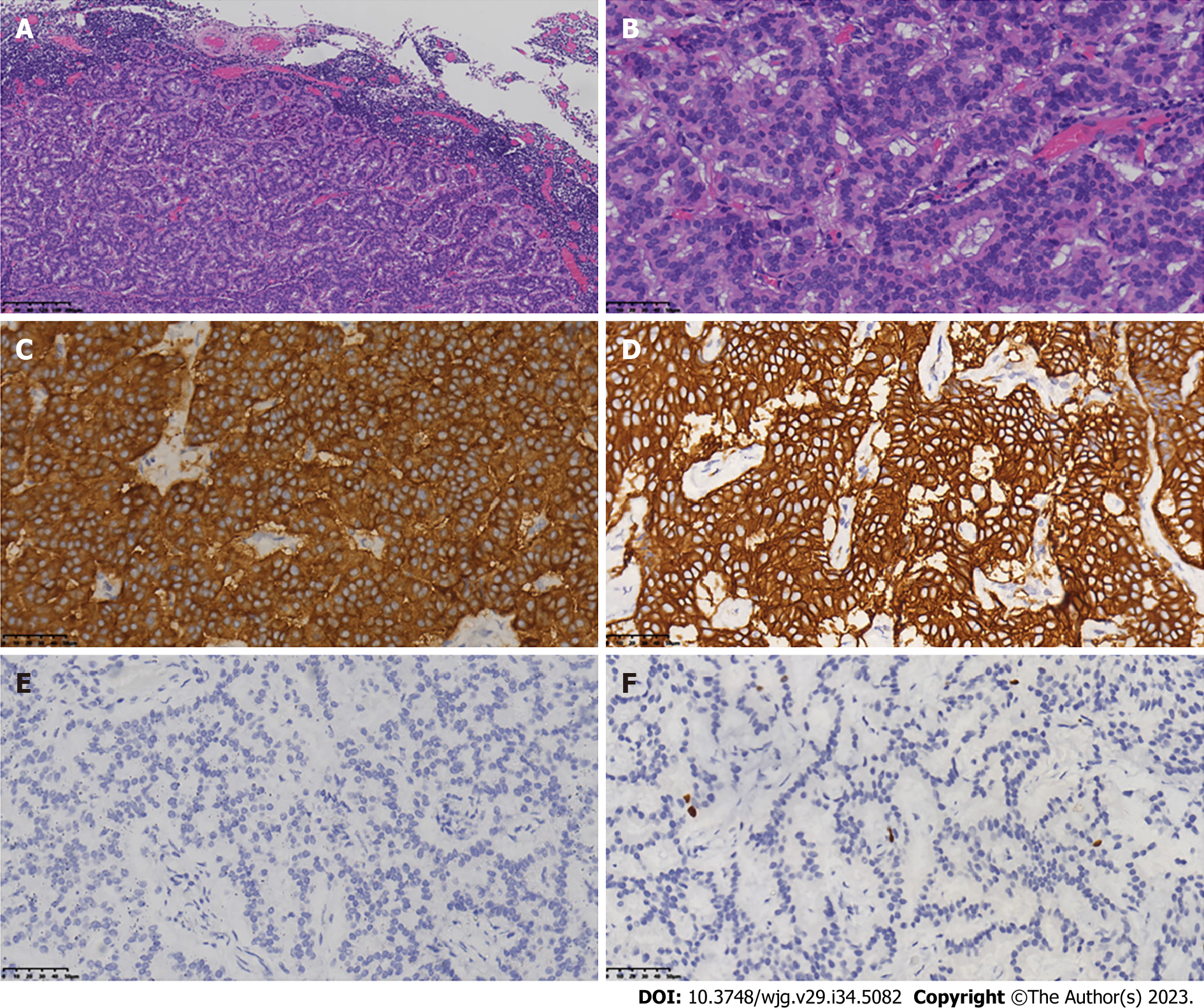
Figure 4 Tumor metastasis was observed in the peri-intestinal lymph nodes (4/15).
A and B: Laparoscopic low anterior resection with total mesorectal excision was performed 5 mo after diagnosis of the rectal lesions. Central lymph nodes were selected from the lesion for hematoxylin–eosin staining to evaluate the metastasis of neuroendocrine cells (A: 100 ×, B: 400 ×); C-E: Immunohistochemical detection to evaluate the expression and distribution of neuroendocrine markers SSTR2, syn, and CgA in central lymph nodes (400 ×); F: Ki-67 expression in central lymph nodes by immunohistochemistry (400 ×).
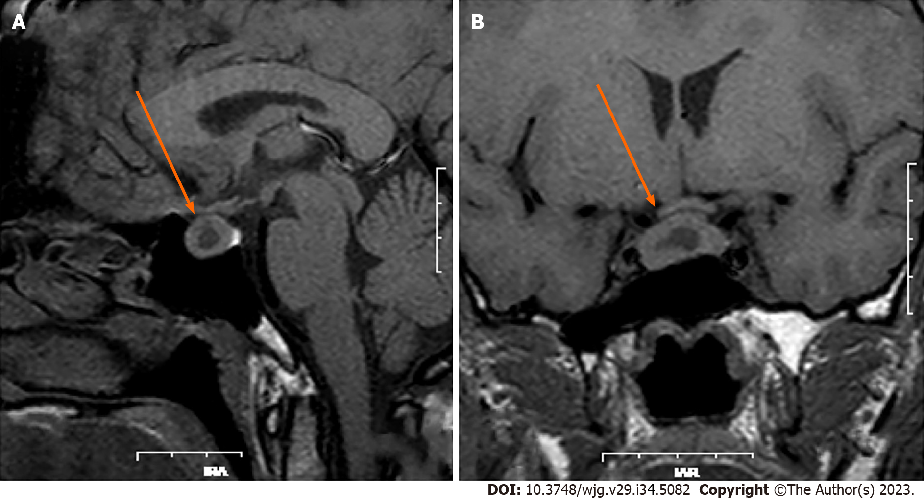
Figure 5 Magnetic resonance imaging for pituitary adenoma.
A: Pretreatment with long-acting octreotide (20 mg); B: Pretreatment with long-acting octreotide (20 mg) for 28 d.
- Citation: Li JY, Chen J, Liu J, Zhang SZ. Simultaneous rectal neuroendocrine tumors and pituitary adenoma: A case report and review of literature. World J Gastroenterol 2023; 29(34): 5082-5090
- URL: https://www.wjgnet.com/1007-9327/full/v29/i34/5082.htm
- DOI: https://dx.doi.org/10.3748/wjg.v29.i34.5082


