Copyright
©The Author(s) 2023.
World J Gastroenterol. May 21, 2023; 29(19): 2992-3002
Published online May 21, 2023. doi: 10.3748/wjg.v29.i19.2992
Published online May 21, 2023. doi: 10.3748/wjg.v29.i19.2992
Figure 1 The procedure of the experimental group.
A: The peritoneum was cut at the lowest point of the peritoneal reflection to enter the anterior rectal space; B: After the incision of the peritoneal reflection, a space can be seen, in which can we easily free the anterior rectal wall. This space is considered the rectovaginal space; C: No other fascial structure was present between the fascia propria of the rectum and the adventitia of the vagina, and these two fascial structures could be pushed away from each other by an ultrasonic knife through blunt separation. ADV: Adventitia of the vagina; FPR: Fascia propria of the rectum; MR: Mesorectum; PR: Peritoneal reflection; R: Rectum; V: Vagina; ARS: Anterior rectal space.
Figure 2 The procedure of the control group.
A: The peritoneum was cut 0.5-1 cm above the peritoneal reflection. The peritoneal reflection was slightly white, and its texture was different from the texture of other structures during the operation; B: The cutting plane was between the vaginal muscle layer and the adventitia. The vaginal adventitia was closely adherent to the muscle layer; C: Bleeding occurred after stripping the vaginal adventitia from the muscle, and hemostasis was performed. ADV: Adventitia of the vagina; FPR: Fascia propria of the rectum; MR: Mesorectum; MUS: Muscle; PR: Peritoneal reflection; R: Rectum; V: Vagina.
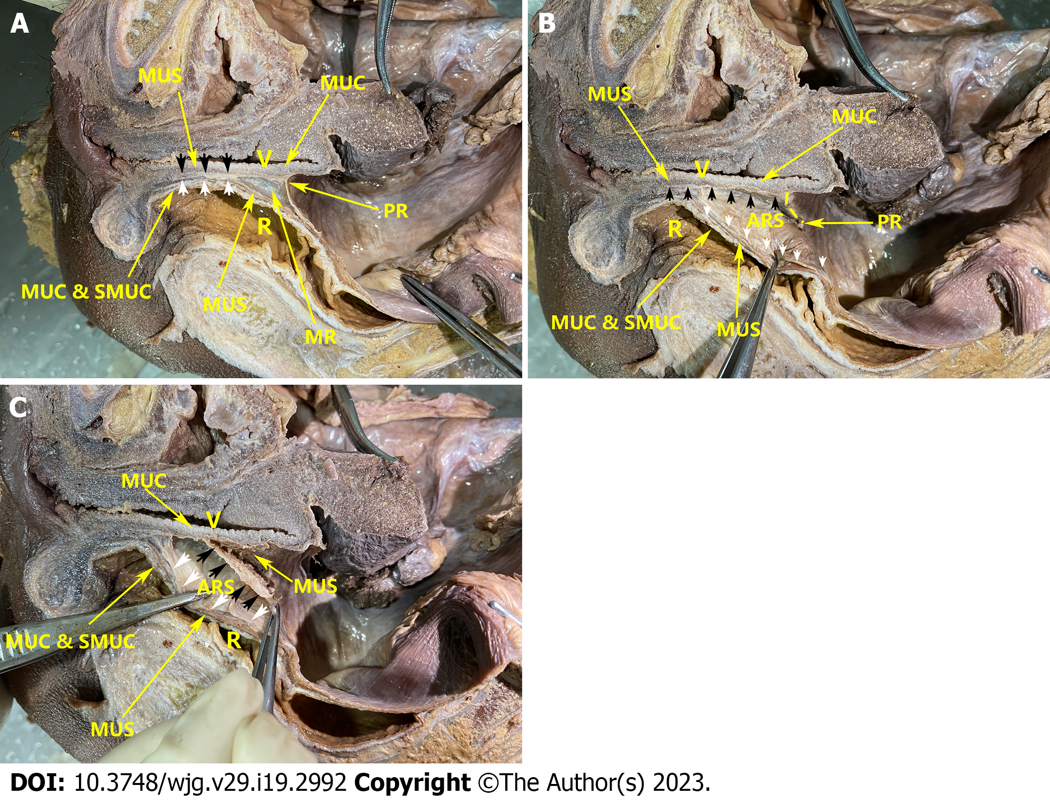
Figure 3 Gross anatomy.
A: Observation of the female pelvis; B: The procedure of the experimental group; C: The procedure of the control group. Black arrow: Adventitia of the vagina; white arrow: Fascia propria of the rectum. ARS: Anterior rectal space; MR: Mesorectum; MUC: Mucosa; MUS: Muscle; PR: Peritoneal reflection; R: Rectum; SMUC: Submucosa; V: Vagina; FPR: Fascia propria of the rectum; MR: Mesorectum.
Figure 4 Pathological section of the rectovaginal structure.
Pathological section showing the absence of an obvious fascia-like structure between the fascia propria of the rectum and the adventitia of the vagina. ADV: Adventitia of the vagina; FPR & MR: Fascia propria of the rectum and mesorectum; MUCV: Mucosa of the vagina; MUSR: Muscle of the rectum; MUSV: Muscle of the vagina; SMR: Submucosa of the rectum; MUCR: Mucosa of the rectum.
- Citation: Jin W, Yang J, Li XY, Wang WC, Meng WJ, Li Y, Liang YC, Zhou YM, Yang XD, Li YY, Li ST. Where is the optimal plane to mobilize the anterior rectal wall in female patients undergoing total mesorectal excision? World J Gastroenterol 2023; 29(19): 2992-3002
- URL: https://www.wjgnet.com/1007-9327/full/v29/i19/2992.htm
- DOI: https://dx.doi.org/10.3748/wjg.v29.i19.2992


