Copyright
©The Author(s) 2022.
World J Gastroenterol. May 28, 2022; 28(20): 2227-2242
Published online May 28, 2022. doi: 10.3748/wjg.v28.i20.2227
Published online May 28, 2022. doi: 10.3748/wjg.v28.i20.2227
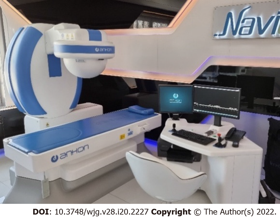
Figure 1 Robotic C-arm, investigation table, and computer workstation for magnetically controlled capsule endoscopy.
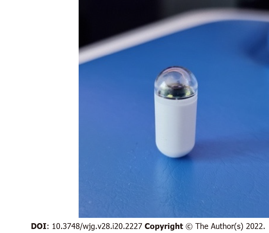
Figure 2 Magnetic capsule endoscope (NaviCam).
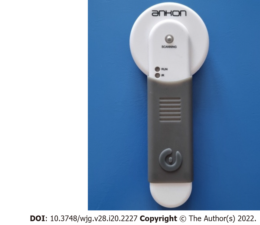
Figure 3 Capsule locator device.
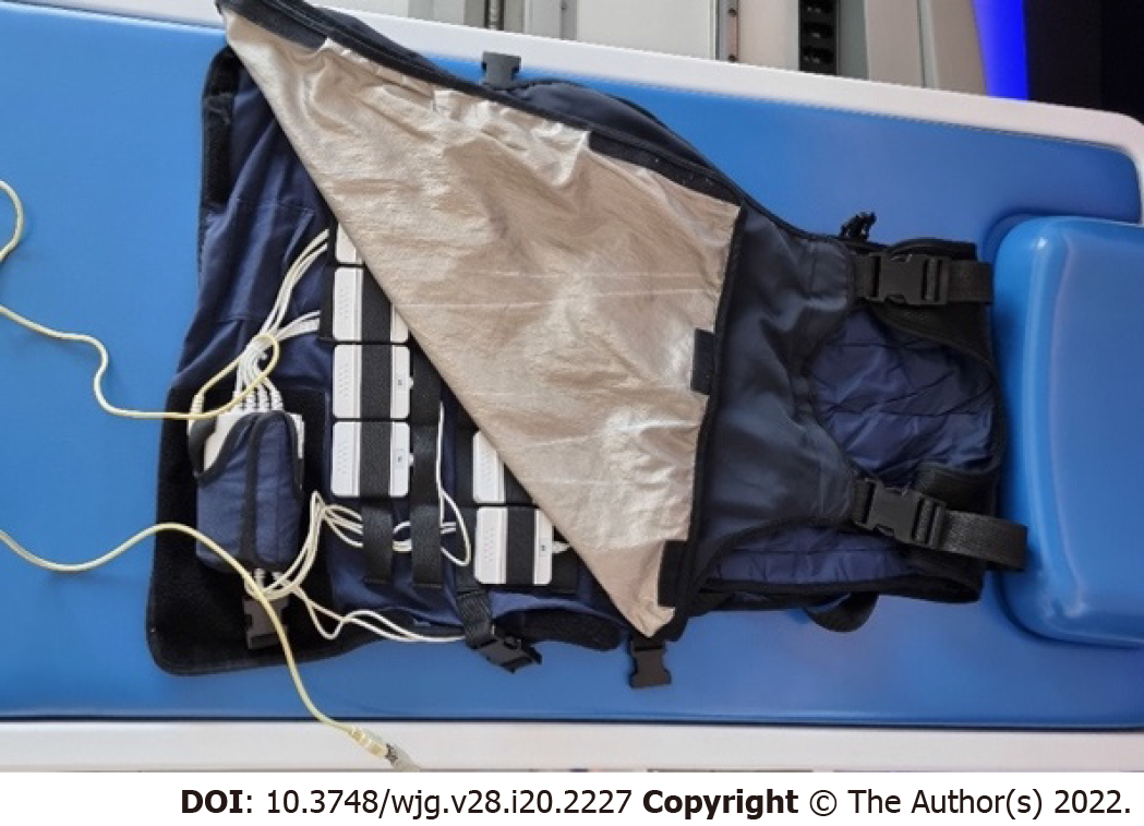
Figure 4 Data recorder.
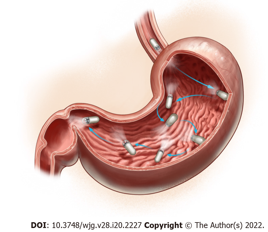
Figure 5 Different magnetically controlled capsule endoscopy stations and capsule camera orientations defined to achieve a complete gastric mucosal surface visualization and mapping (created by Zoltán Tóbiás, MD).
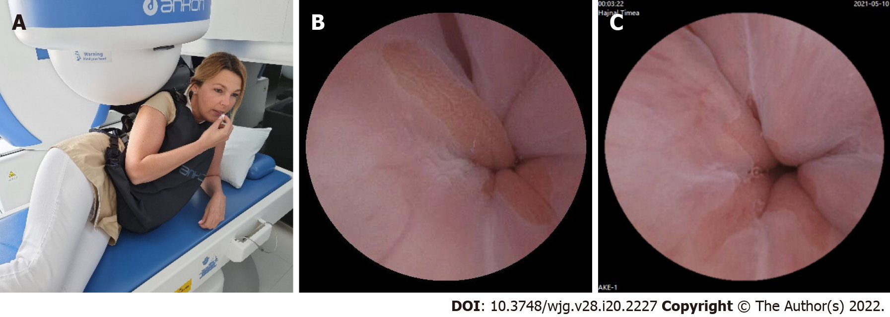
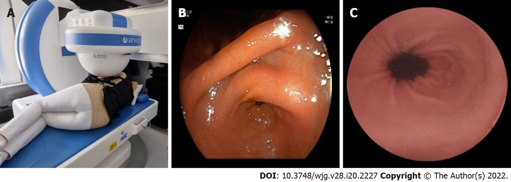
Figure 6 The capsule swallowed by the patients in the left lateral decubitus position.
A: Patients and ball magnet position; B and C: Pictures of the Z-line by CE from our database.
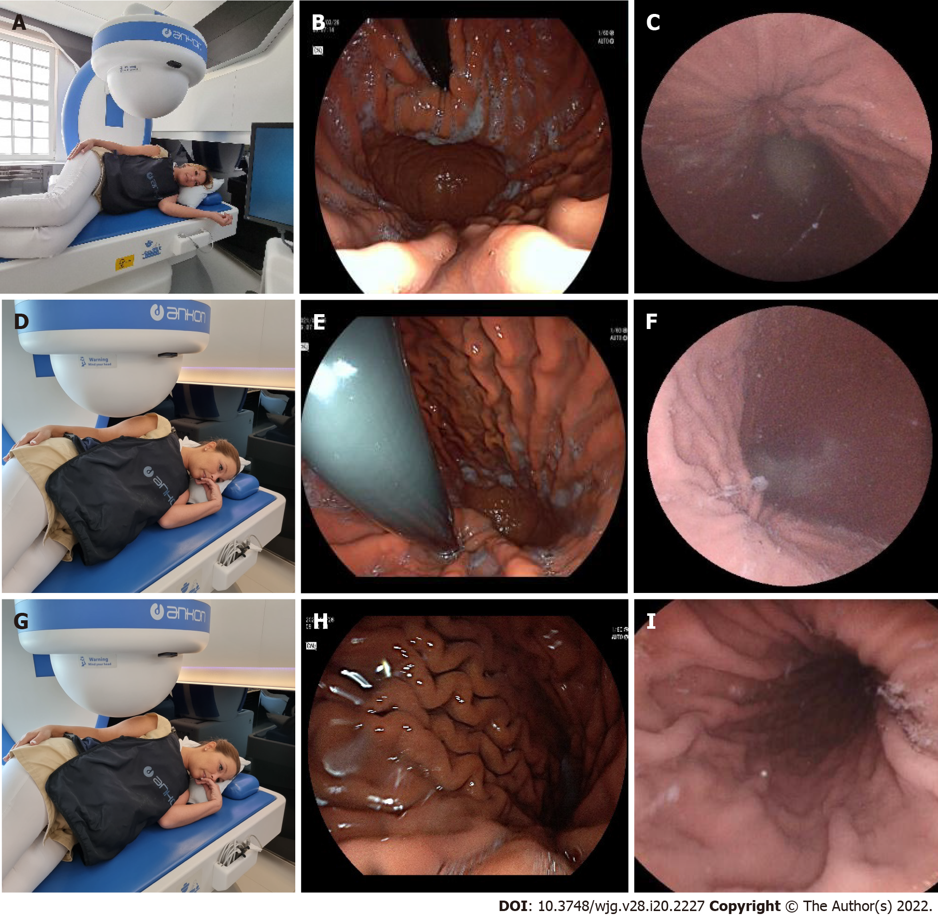
Figure 7 Stations I-III at the left lateral decubitus position.
A: Patients’ position (Station I); B: Cardia by a gastroscope (Station I); C: Cardia by CE (Station I); D: Patients’ position (Station II); E: Cardia by a gastroscope (Station II); F: Cardia by CE (Station II); G: Patients’ position (Station III); H: Corpus by a gastroscope (Station III); I: Corpus by a capsule (Station III).
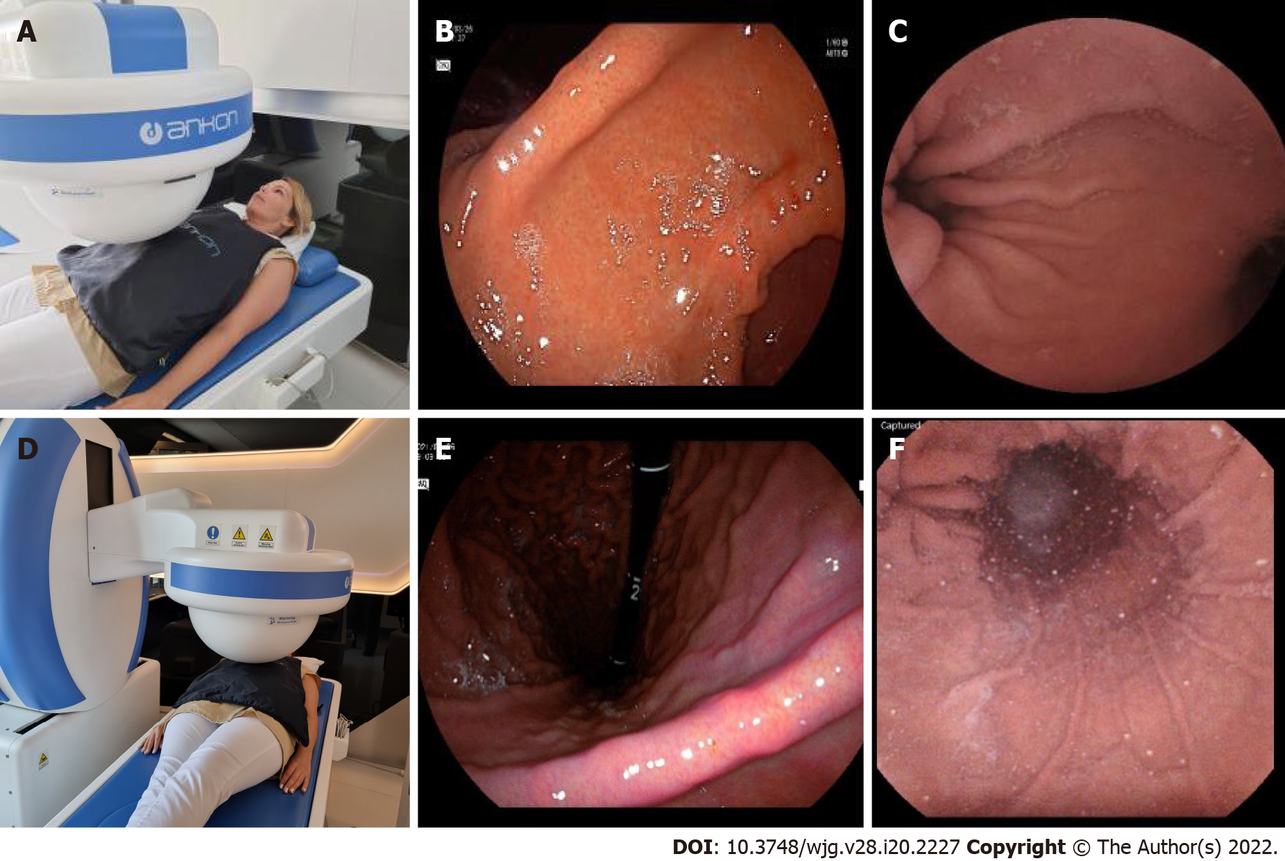
Figure 8 Stations IV and V in the supine position.
A: Patients’ position (Station IV); B: Angular incisure by a gastroscope (Station IV); C: Angular incisure by CE (Station IV); A: Patients’ position (Station V); B: Angular incisure by a gastroscope (Station V); C: Angular incisure by CE (Station V).
Figure 9 Station at the right lateral decubitus patient position.
A: Patients’ position (Station VI); B: Antrum canal of stomach by gastroscope (Station VI); C: Antrum canal of stomach by CE (Station VI).
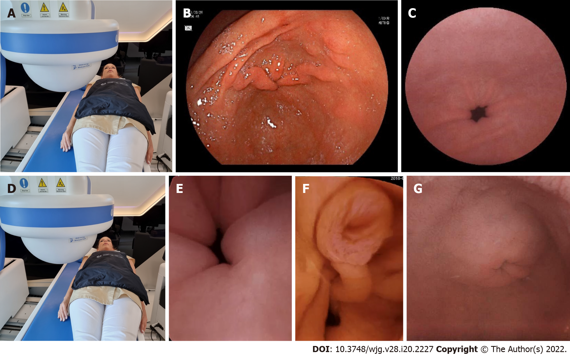
Figure 10 Stations VII and VIII in the supine position.
A: Patient’s position (Station VII); B: Antrum canal of stomach by gastroscope (Station VII); C: Antrum canal of stomach by CE (Station VII); D: Patient’s position (Station VIII); E: Pyloric ring (Station VIII); F: Ampulla of Vater (Station VIII); G: Pylorus from the duodenal bulb with a capsule (Station VIII).
- Citation: Szalai M, Helle K, Lovász BD, Finta Á, Rosztóczy A, Oczella L, Madácsy L. First prospective European study for the feasibility and safety of magnetically controlled capsule endoscopy in gastric mucosal abnormalities. World J Gastroenterol 2022; 28(20): 2227-2242
- URL: https://www.wjgnet.com/1007-9327/full/v28/i20/2227.htm
- DOI: https://dx.doi.org/10.3748/wjg.v28.i20.2227


