Copyright
©The Author(s) 2020.
World J Gastroenterol. Nov 7, 2020; 26(41): 6361-6377
Published online Nov 7, 2020. doi: 10.3748/wjg.v26.i41.6361
Published online Nov 7, 2020. doi: 10.3748/wjg.v26.i41.6361
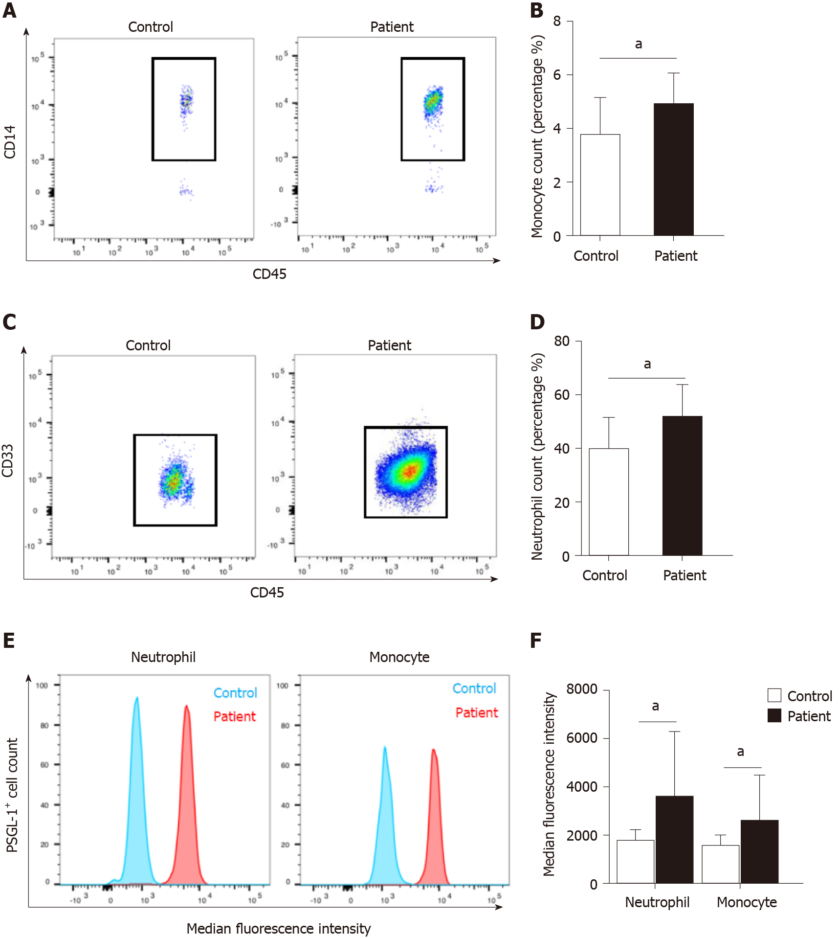
Figure 1 Increased expression of P-selectin glycoprotein ligand 1 on neutrophils and monocytes from patients with acute pancreatitis.
A: Flow cytometry for CD14-positive cells to detect monocytes in the peripheral blood of patients (n = 21) with acute pancreatitis (AP) or normal control subjects (n = 8); B: Boxplot showing quantification of monocytes; C: Flow cytometry for CD33-positive cells to detect neutrophils in the peripheral blood of patients (n = 21) with AP or normal control subjects (n = 8); D: Boxplot showing quantification of neutrophils; E: Fluorescence intensity of P-selectin glycoprotein ligand 1 (PSGL-1) on neutrophils and monocytes; F: Boxplot showing quantification of fluorescence intensity of PSGL-1 on neutrophils and monocytes. aP < 0.05, Student's t-test. PSGL-1: P-selectin glycoprotein ligand 1.
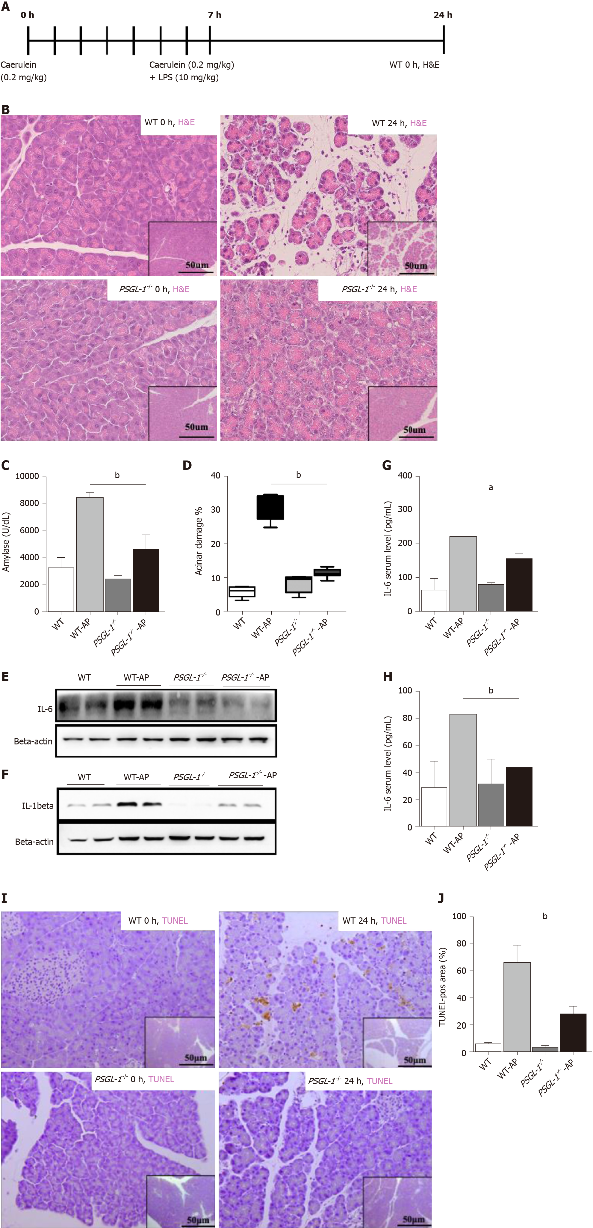
Figure 2 P-selectin glycoprotein ligand 1 deficiency alleviates caerulein-mediated inflammatory response and acinar damage.
A: Schematic diagram of the induction of acute pancreatitis (AP) showing the frequency of caerulein and lipopolysaccharide injections as well as the sampling time points; B: Hematoxylin-eosin staining of the pancreas of wild-type (upper panels) and P-selectin glycoprotein ligand 1 (PSGL-1)-/- (lower panels) mice 0 h and 24 h after treatment with caerulein; C: Boxplots showing the expression of amylase (n = 4); D: Boxplots showing quantification of acinar cell damage (n = 4); E: Expression of IL-6 in the pancreas of wild-type and PSGL-1-/- mice; F: Expression of IL-1beta in the pancreas of wild-type and PSGL-1-/- mice; G: Expression of IL-6 in sera of wild-type and PSGL-1-/- mice (n = 4); H: Expression of IL-1beta in sera of wild-type and PSGL-1-/- mice(n = 4); I: Transferase-mediated dUTP-biotin nick end labeling (TUNEL) assay for apoptosis in the pancreas; J: Boxplots showing quantification of the TUNEL assays (n = 4). aP < 0.05; bP < 0.001, Student's t-test. H&E: Hematoxylin-eosin; TUNEL: Transferase-mediated dUTP-biotin nick end labeling; PSGL-1: P-selectin glycoprotein ligand 1; AP: Acute pancreatitis; WT: Wild type.
Figure 3 P-selectin glycoprotein ligand 1 deficiency attenuates caerulein-mediated leukocyte infiltration in the pancreas.
A: Immunohistochemistry of pancreatic tissue sections for F4/80 (monocytes/macrophages); B: Immunohistochemistry of pancreatic tissue sections for myeloperoxidase (MPO) (neutrophils); C: Boxplots showing quantification of F4/80-positive monocytes/macrophages in the pancreas (n = 4); D: Boxplots showing quantification of MPO-positive neutrophils in the pancreas (n = 4); E: Immunohistochemistry of pancreatic tissue sections stained for CD3 (T cells); F: Immunohistochemistry of pancreatic tissue sections stained for CD45R (B cells); G: Boxplots showing quantification of CD3-positive T cells in the pancreas (n = 4); H: Boxplots showing quantification of CD45R-positive B cells in the pancreas (n = 4); aP < 0.001, bP < 0.01, Student's t-test. PSGL-1: P-selectin glycoprotein ligand 1; MPO: Myeloperoxidase; WT: Wild type; AP: Acute pancreatitis.
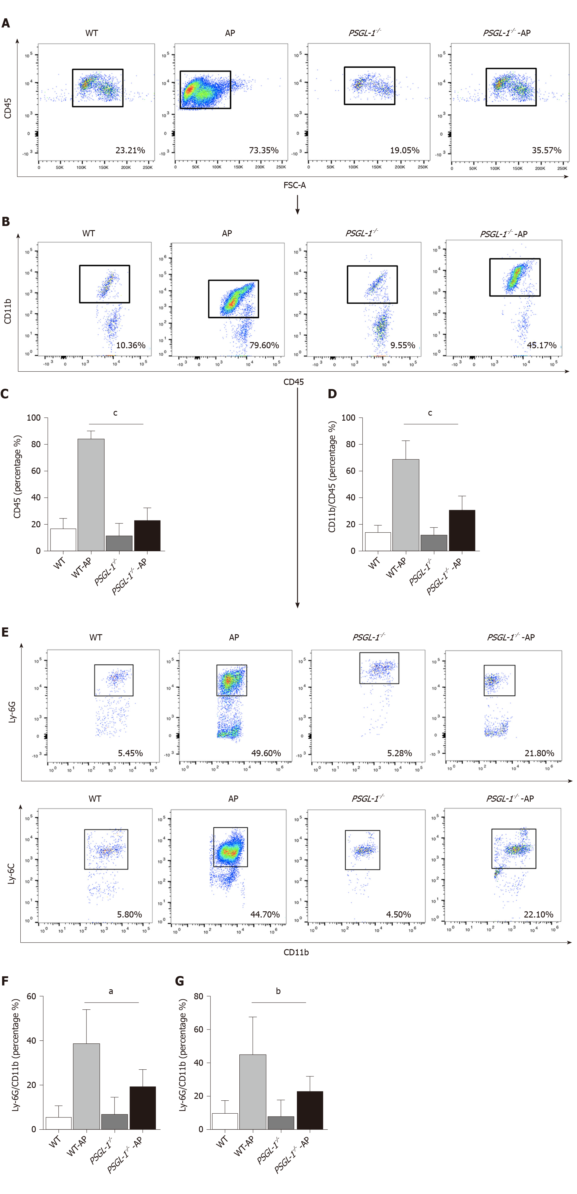
Figure 4 P-selectin glycoprotein ligand 1 deficiency attenuates the number of circulating leukocytes.
A: Flow cytometry for CD45-positive cells to detect immune cells in the serum; B: Flow cytometry for CD11b-positive cells to detect myeloid cells in the serum; C: Boxplots showing quantification of immune cells in the serum (n = 4); D: Boxplots showing quantification of myeloid cells in the serum (n = 4); E: Flow cytometry for Ly-6G-positive cells and Ly-6C-positive cells to detect neutrophils and monocytes, respectively, in the serum; F: Boxplots showing quantification of neutrophils in the serum (n = 4); G: Boxplots showing quantification of monocytes in the serum (n = 4). aP < 0.05; bP < 0.01, cP < 0.001, Student's t-test. PSGL-1: P-selectin glycoprotein ligand 1; WT: Wild type; AP: Acute pancreatitis.
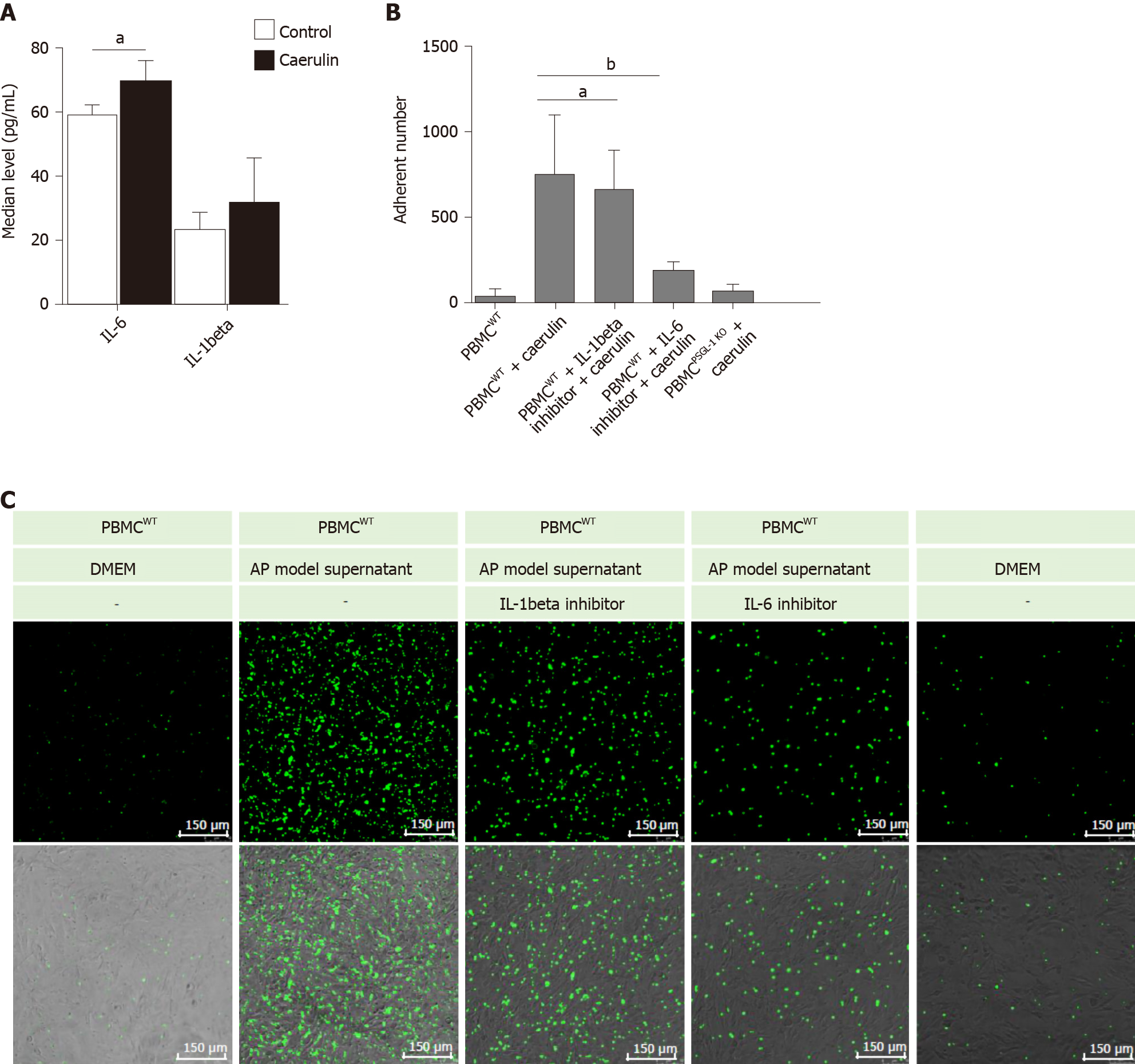
Figure 5 P-selectin glycoprotein ligand 1 deficiency alleviates caerulein-induced leukocyte-endothelial cell adhesion.
A: Enzyme-linked immunosorbent assays for IL-6 and IL-1beta in the supernatants of AR42J cells cultured in caerulein-containing medium (n = 4); B: Boxplots showing the quantification of adhered peripheral blood mononuclear cells (PBMCs) (n = 4); C: The number of PBMCs that adhered to Bend.3 cells cocultured under different conditions. Bend. 3 cells were cocultured with PBMCs from wild-type or PSGL-1-/- mice and stained with Green 5-chloromethylformacein diacetate under different conditions. Fluorescent signals were captured using a confocal microscope (Leica, 400 ×). Green represents the number of adhered PBMCs. aP < 0.05; bP < 0.01, Student's t-test. PSGL-1: P-selectin glycoprotein ligand 1; WT: Wild type; PBMCs: Peripheral blood mononuclear cells; DMEM: Dulbecco's modified Eagle's medium; AP: Acute pancreatitis.
- Citation: Zhang X, Zhu M, Jiang XL, Liu X, Liu X, Liu P, Wu XX, Yang ZW, Qin T. P-selectin glycoprotein ligand 1 deficiency prevents development of acute pancreatitis by attenuating leukocyte infiltration. World J Gastroenterol 2020; 26(41): 6361-6377
- URL: https://www.wjgnet.com/1007-9327/full/v26/i41/6361.htm
- DOI: https://dx.doi.org/10.3748/wjg.v26.i41.6361


