Copyright
©The Author(s) 2020.
World J Gastroenterol. Oct 7, 2020; 26(37): 5731-5744
Published online Oct 7, 2020. doi: 10.3748/wjg.v26.i37.5731
Published online Oct 7, 2020. doi: 10.3748/wjg.v26.i37.5731
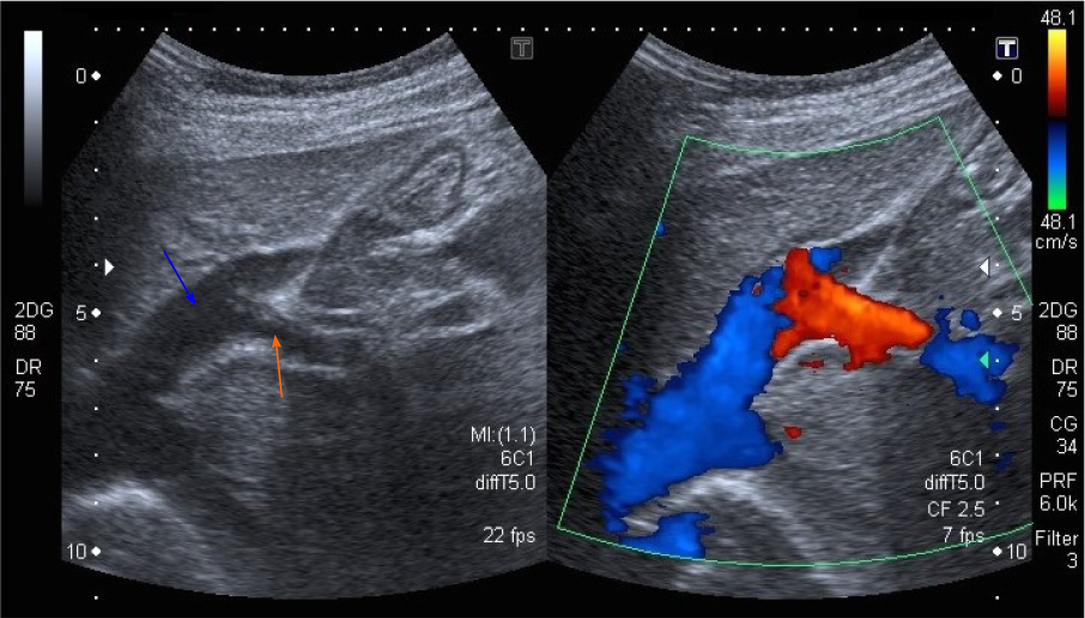
Figure 1 Abdominal ultrasound with Doppler color flow (case 1).
An anomalous vascular connection between the left hepatic vein (blue arrow) and left portal vein (orange arrow) is seen.
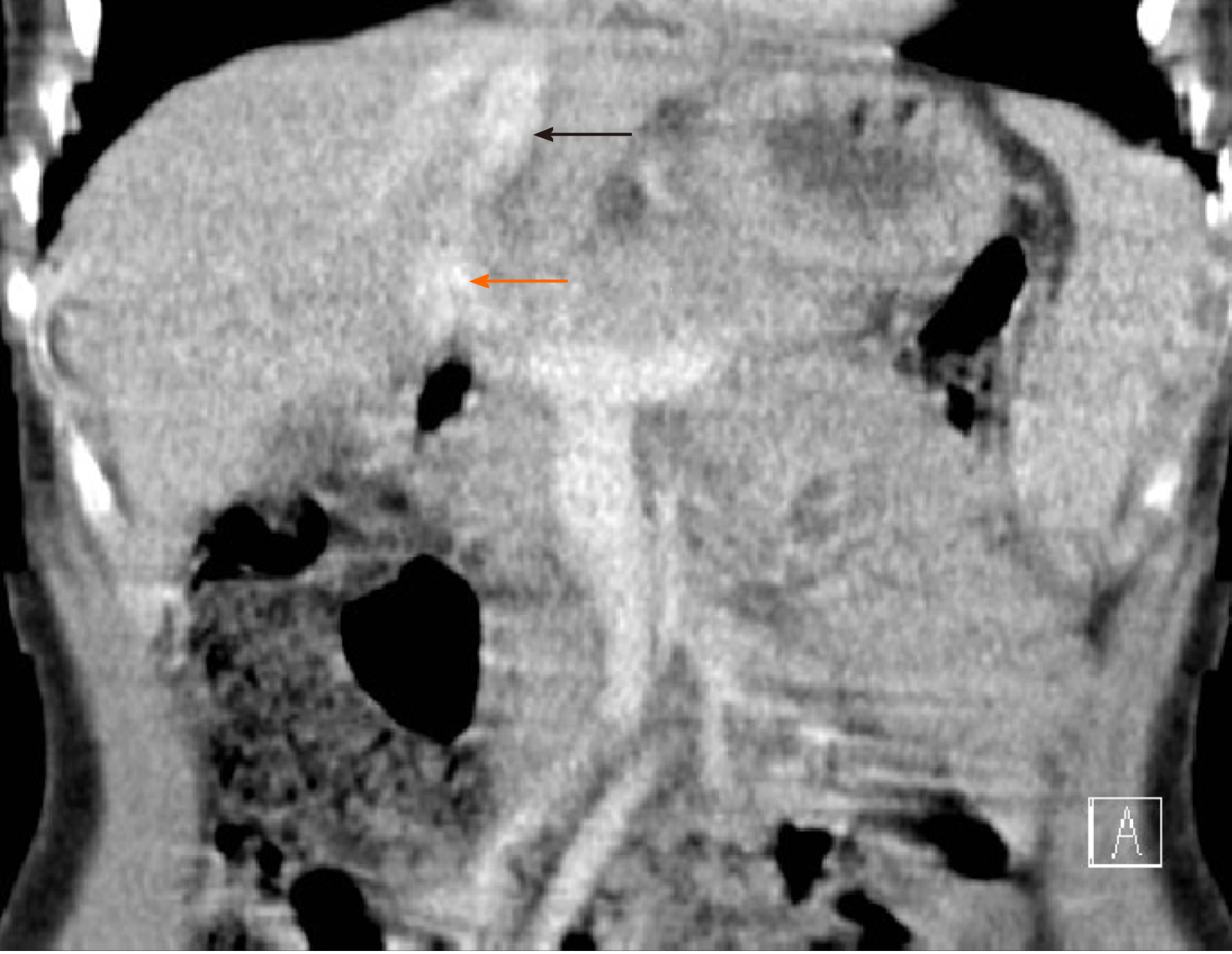
Figure 2 Abdominal computed tomography image of the intrahepatic portosystemic shunt (case 1).
The left portal vein (orange arrow) flows directly into the left hepatic vein (black arrow).
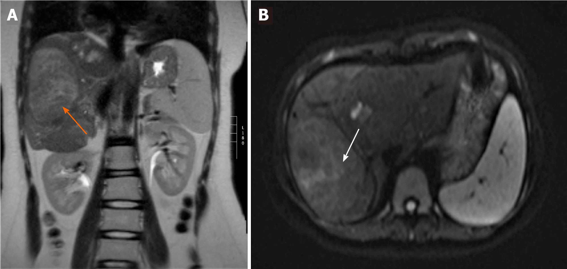
Figure 3 Abdominal magnetic resonance imaging scan demonstrating a lesion in the right liver lobe (case 5).
A: The lesion (orange arrow) is slightly hyperintense compared to the sorrounding liver tissue on coronal T2-weighted imaging; B: The same lesion (white arrow) has no restriction of diffusion on axial diffusion-weighted imaging (DWI) sequence - a benign lesion.
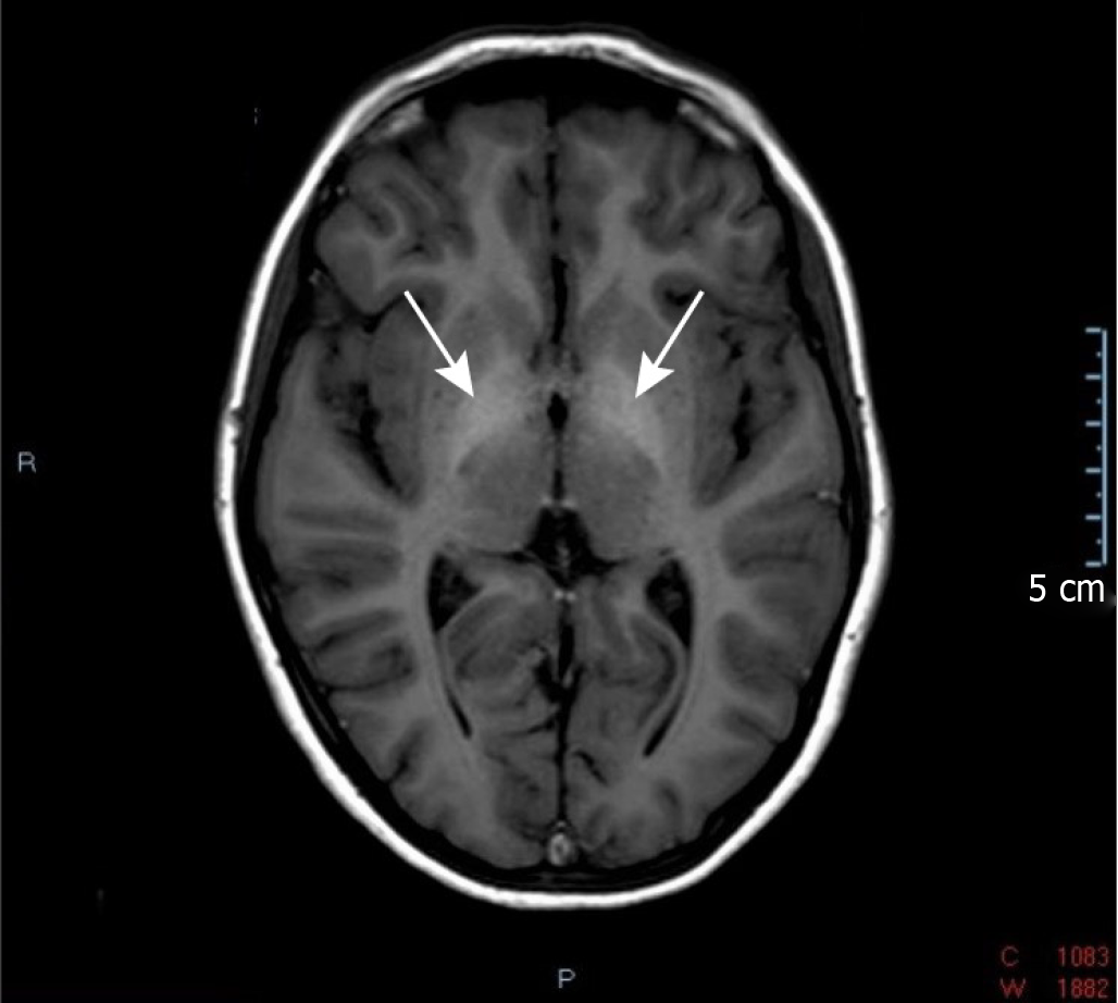
Figure 4 Axial T1-weighted brain magnetic resonance imaging scan (case 5).
Arrows indicate symmetrical T1-hyperintensity involving the bilateral globus pallidus.
- Citation: Peček J, Fister P, Homan M. Abernethy syndrome in Slovenian children: Five case reports and review of literature. World J Gastroenterol 2020; 26(37): 5731-5744
- URL: https://www.wjgnet.com/1007-9327/full/v26/i37/5731.htm
- DOI: https://dx.doi.org/10.3748/wjg.v26.i37.5731


