Copyright
©The Author(s) 2019.
World J Gastroenterol. Mar 21, 2019; 25(11): 1421-1431
Published online Mar 21, 2019. doi: 10.3748/wjg.v25.i11.1421
Published online Mar 21, 2019. doi: 10.3748/wjg.v25.i11.1421
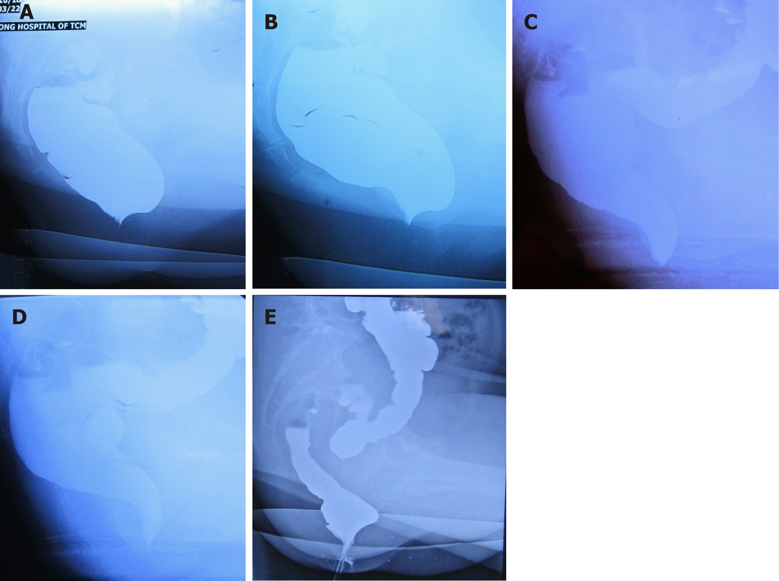
Figure 1 Defecography images for rectocele patients.
A: Preoperative defecography for a patient in group A; B: Preoperative defecography for a patient in group B; C: Postoperative defecography for the patient in group A; D: Postoperative defecography for the patient in group B; E: Defecography image of rectocele recurrence.
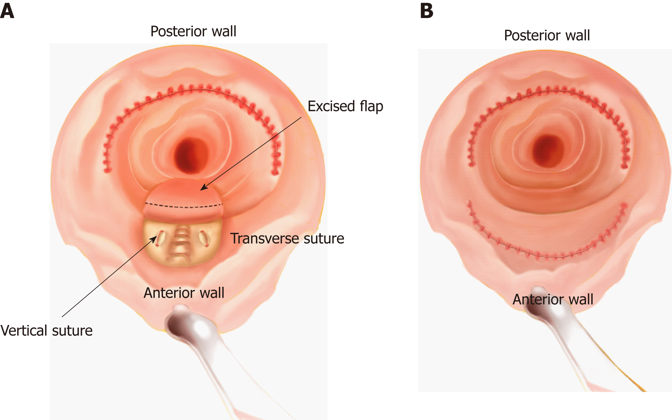
Figure 2 Sketch of the operation.
A: Khubchandani’s procedure with stapled posterior rectal wall resection. Stapled posterior wall repair is performed. Subsequently, a U-shape flap is freed, and three to five transverse and two vertical sutures are made in the muscular layer. Then, most part of the U-shape flap is excised and sutured; B: Stapled transanal rectal resection. Stapled rectal wall repair is performed in both the anterior and posterior wall.
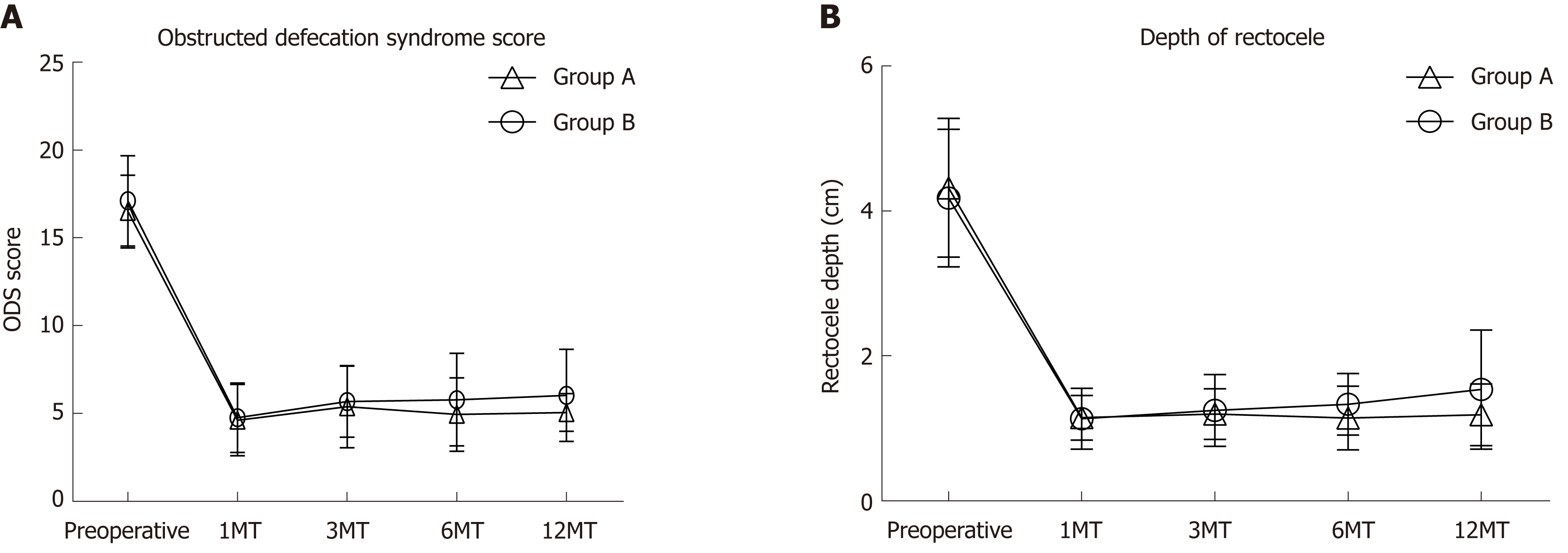
Figure 3 Preoperative and postoperative obstructed defecation syndrome scores and rectocele depth.
A: Obstructed defecation syndrome score. There was a statistical difference between groups A and B at 1 year after operation; B: Rectocele depth. There was a statistical difference between groups A and B at 1 year after operation. ODS: Obstructed defecation syndrome; 1MT: 1 mo after operation; 3MT: 3 mo after operation; 6MT: 6 mo after operation; 12MT: 1 year after operation.
- Citation: Shao Y, Fu YX, Wang QF, Cheng ZQ, Zhang GY, Hu SY. Khubchandani’s procedure combined with stapled posterior rectal wall resection for rectocele. World J Gastroenterol 2019; 25(11): 1421-1431
- URL: https://www.wjgnet.com/1007-9327/full/v25/i11/1421.htm
- DOI: https://dx.doi.org/10.3748/wjg.v25.i11.1421


