Copyright
©The Author(s) 2018.
World J Gastroenterol. Sep 28, 2018; 24(36): 4178-4185
Published online Sep 28, 2018. doi: 10.3748/wjg.v24.i36.4178
Published online Sep 28, 2018. doi: 10.3748/wjg.v24.i36.4178
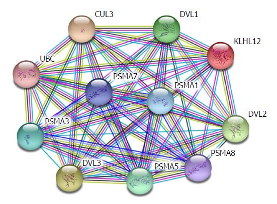
Figure 1 Interaction between WNT-related genes and collagen-related genes.
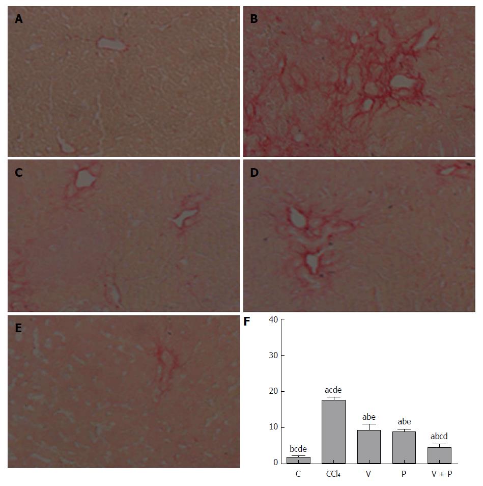
Figure 2 Sirius red staining.
A: Control (C) group; B: CCl4 group; C: Vitamin D (V) group; D: Puerarin (P) group; E: V + P group (200 × magnification); F: Statistical graph of positive Sirius-red-stained areas (in the comparison of the mean ± SD of various groups, differences are statistically significant when P < 0.05, n = 10. a, b, c, d, and e represent statistical differences vs C, CCl4, V, P, and V + P groups, respectively).
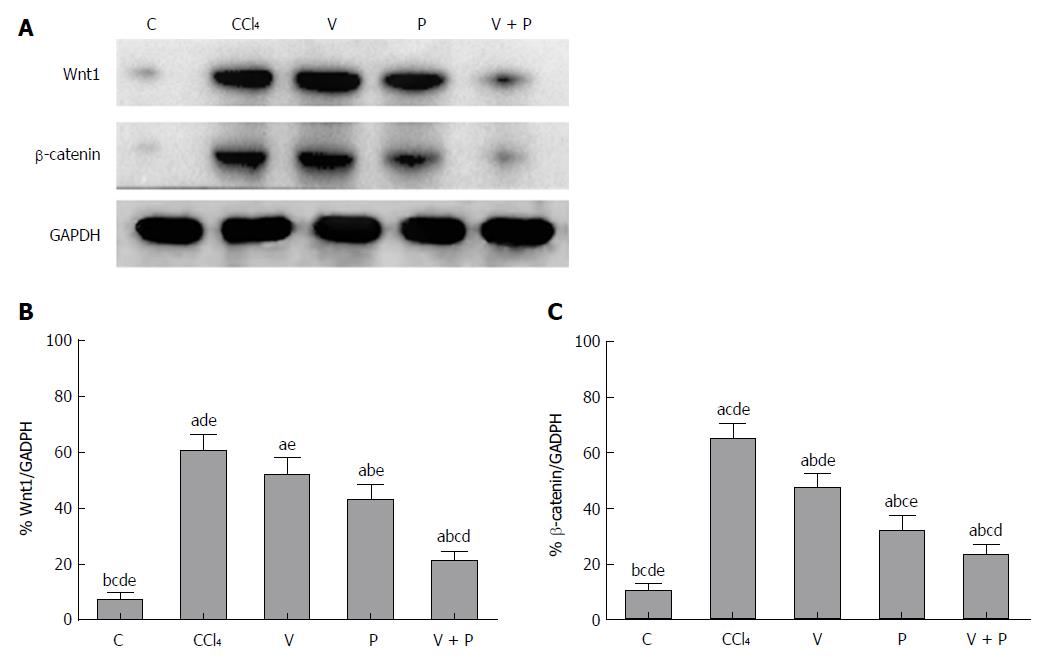
Figure 3 The protein levels of Wnt1 and β-catenin of liver tissues in rats in the CCl4 group and the medicated groups were higher than the Control (C) group.
A: Detection of protein levels of Wnt1, and β-catenin using western blot assay; B: Grey analysis of Wnt1; C: Grey analysis of β-catenin [in the comparison of mean ± standard deviation (SD) of various groups, differences are statistically significant when P < 0.05, n = 10. In addition, a, b, c, d, and e represent statistical differences vs C, CCl4, V, P, and V + P groups, respectively]. C: Control; V: Vitamin; P: Puerarin.
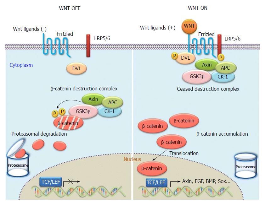
Figure 4 Overview of WNT/β-catenin signalling.
Without WNT signalling (“WNT OFF”), the destruction complex phosphorylates cytosolic β-catenin, and phosphorylated β-catenin is recognized and degraded by the proteasomes. With active WNT signalling (“WNT ON”), the function of the “destruction complex” is inhibited to phosphorylate cytosolic β-catenin. Then unphosphorylated β-catenin accumulates in the cytosol, translocates into the nucleus, and activates WNT target gene expression, such as the targets of the T-cell factor and lymphoid enhancer factor-1 (TCF/LEF1) family of transcription factors.
- Citation: Huang GR, Wei SJ, Huang YQ, Xing W, Wang LY, Liang LL. Mechanism of combined use of vitamin D and puerarin in anti-hepatic fibrosis by regulating the Wnt/β-catenin signalling pathway. World J Gastroenterol 2018; 24(36): 4178-4185
- URL: https://www.wjgnet.com/1007-9327/full/v24/i36/4178.htm
- DOI: https://dx.doi.org/10.3748/wjg.v24.i36.4178


