Copyright
©The Author(s) 2017.
World J Gastroenterol. Jun 21, 2017; 23(23): 4293-4302
Published online Jun 21, 2017. doi: 10.3748/wjg.v23.i23.4293
Published online Jun 21, 2017. doi: 10.3748/wjg.v23.i23.4293
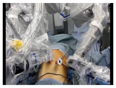
Figure 1 Robotic docking.
Figure 2 Double loop reconstruction method.
A: 1 step: E-J anastomosis; B: 2 step: J-J anastomosis using the second loop; C: 3 step: interruption of continuity between the two anastomoses.
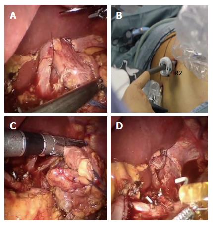
Figure 3 Division of the esophagus.
A: After fixing the esophagus to the diaphragm pillars; B: An articulated mechanical linear stapler is introduced by the assistant through a 12 mm trocar; C and D: The esophagus is sectioned and closed.
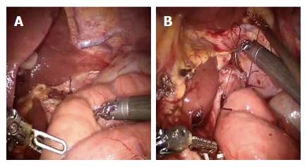
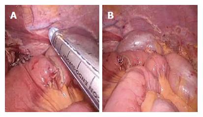
Figure 4 A first loop of small bowel is identified and brought antecolic (A), two stiches are placed to pair it with the esophagus (B).
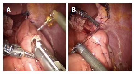
Figure 5 Esophagus and jejunum are opened (A), the posterior layer (B) is first performed followed by the anterior layer (C and D).
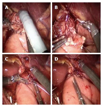
Figure 6 Second loop is identified at 40 cm from the E-J anastomosis, along the alimentary limb, and brought up to the first anastomosis on its left side through a mechanical stapler (A), a side to side jejunojejunal anastomosis is created between the second loop and the biliary limb of the first loop (B).
Figure 7 Two anastomoses are easily interrupted by firing the stapler (A and B).
- Citation: Parisi A, Ricci F, Gemini A, Trastulli S, Cirocchi R, Palazzini G, D’Andrea V, Desiderio J. New totally intracorporeal reconstructive approach after robotic total gastrectomy: Technical details and short-term outcomes. World J Gastroenterol 2017; 23(23): 4293-4302
- URL: https://www.wjgnet.com/1007-9327/full/v23/i23/4293.htm
- DOI: https://dx.doi.org/10.3748/wjg.v23.i23.4293


