Copyright
©The Author(s) 2016.
World J Gastroenterol. Feb 14, 2016; 22(6): 2142-2148
Published online Feb 14, 2016. doi: 10.3748/wjg.v22.i6.2142
Published online Feb 14, 2016. doi: 10.3748/wjg.v22.i6.2142
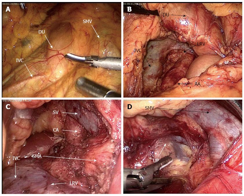
Figure 1 Surgical method - probing.
A: After lifting the transverse mesocolon and exposing the inferior duodenal flexure from its root, the superomedial side of this section is SMV, and the posterolateral side is IVC, and in here, they are the “window” of LPD; B: After lifting the inferior duodenal flexure, freeing along its rear part, entering the Toldt’s gap, revealing the inferior vena cava and left renal vein, and freeing leftwards to reveal the abdominal aorta, the para-abdominal aortic lymph nodes are obtained and sent for the intraoperative frozen section; C: After exposing SMA root from the upper site of LRV and dissecting along its distribution from the rear part, whether tumour invades the SMA or not is explored. The freeing is then continued towards the caput to reveal the VA root and clean its surrounding lymph nodes; D: After revealing the SMV from inferior duodena part, this segment has longer SMV, entirely located within the small bowel mesentery, and has no avascular association with the duodenum. SMV: Superior mesenteric vein; IVC: Inferior vena cava; DU: Duodenum; LPD: Laparoscopic pancreaticoduodenectomy; LRV: Left renal vein; AA: Abdominal aorta; CA: Celiac artery; UP: Uncinate process.
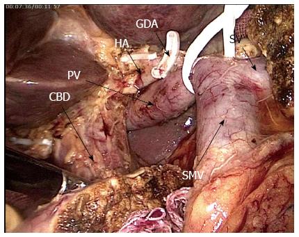
Figure 2 Surgical method - specimen dissection.
After entering the hepatoduodenal ligament, dissecting GDA, and freeing towards the hepatic porta along the surface of hepatic portal vein, it is separated from the hepatic artery and common bile duct. The hepatic artery is fully dissected, and the surrounding fat lymphoid tissues are also dissected. Finally, the common bile duct is dissected. GDA: Gastric-duodenum artery; HA: Hepatic artery; PV: Portal vein; CBD: Common bile duct; SMV: Superior mesenteric vein; SV: Spleen vein.
- Citation: Wang XM, Sun WD, Hu MH, Wang GN, Jiang YQ, Fang XS, Han M. Inferoposterior duodenal approach for laparoscopic pancreaticoduodenectomy. World J Gastroenterol 2016; 22(6): 2142-2148
- URL: https://www.wjgnet.com/1007-9327/full/v22/i6/2142.htm
- DOI: https://dx.doi.org/10.3748/wjg.v22.i6.2142


