Copyright
©The Author(s) 2016.
World J Gastroenterol. Dec 21, 2016; 22(47): 10461-10464
Published online Dec 21, 2016. doi: 10.3748/wjg.v22.i47.10461
Published online Dec 21, 2016. doi: 10.3748/wjg.v22.i47.10461
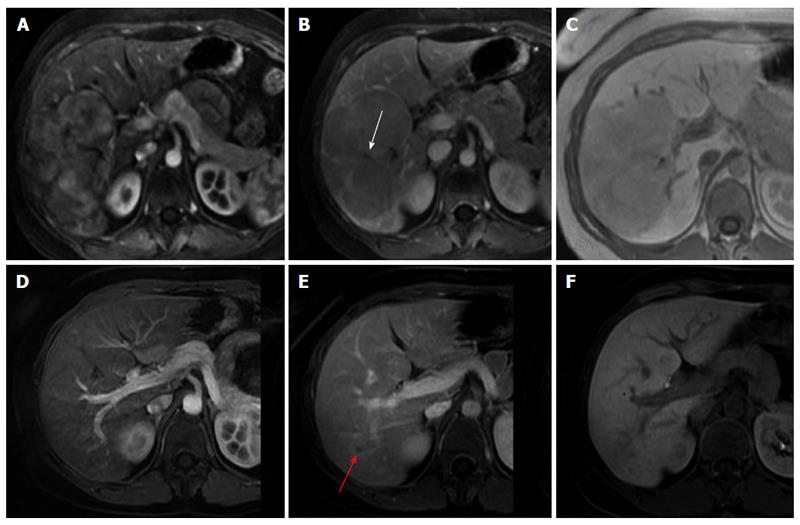
Figure 1 Magnetic resonance imaging showing a typical large focal nodular hyperplasia in 2005 (A-C) with complete spontaneous regression after 7 years of follow-up (D-F).
Gd-BOPTA-enhanced magnetic resonance imaging showed a large hypervascular lesion on arterial phase imaging (A), with iso to hypoenhancement on the delayed venous phase (B), and uptake of Gd-BOPTA on the 2 h delayed hepatobiliary phase (C). Note the central scar inside the lesion (white arrow). In 2012, MRI with the same contrastographic sequences demonstrated complete resolution (D-F), with residual subcentimeter scarring (red arrow).
- Citation: Mamone G, Caruso S, Cortis K, Miraglia R. Complete spontaneous regression of giant focal nodular hyperplasia of the liver: Magnetic resonance imaging evaluation with hepatobiliary contrast media. World J Gastroenterol 2016; 22(47): 10461-10464
- URL: https://www.wjgnet.com/1007-9327/full/v22/i47/10461.htm
- DOI: https://dx.doi.org/10.3748/wjg.v22.i47.10461


