Copyright
©The Author(s) 2016.
World J Gastroenterol. Nov 7, 2016; 22(41): 9222-9228
Published online Nov 7, 2016. doi: 10.3748/wjg.v22.i41.9222
Published online Nov 7, 2016. doi: 10.3748/wjg.v22.i41.9222
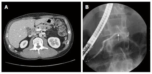
Figure 1 Enhanced abdominal computed tomography scan (A) and endoscopic retrograde cholangiopancreatography image (B) of case 1.
A: The hypovascular tumor in the pancreatic body (arrowheads); B: Disruption of the main pancreatic duct (arrow).
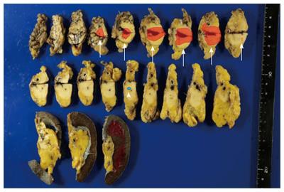
Figure 2 Image of the resected pancreas from case 1.
The resected specimen showed a 35 mm × 18 mm tumor in the pancreatic body (arrows). Pathological findings revealed a small lesion at a 20-mm distant to the tail side from the main lesion (arrowhead).
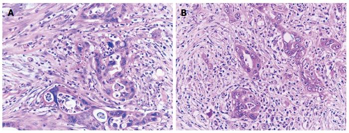
Figure 3 Microscopic findings of the main lesion (A) and the daughter lesion (B) of case 1.
A: Tumor cells form irregular glands with marked infiltration of neutrophils. Hematoxylin and eosin staining. Objective magnification, × 40; B: The microscopic lesion of the pancreatic tail demonstrated similar morphology to the main lesion. Hematoxylin and eosin staining. Objective magnification, × 40.
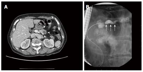
Figure 4 Enhanced abdominal computed tomography scan (A) and endoscopic retrograde cholangiopancreatography image (B) of case 2.
A: The hypovascular tumor in the pancreatic body (arrowheads); B: Dilation and disruption of the main pancreatic duct (arrows).
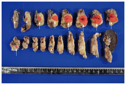
Figure 5 Image of the resected pancreas from case 2.
The resected specimen showed a 32 mm × 20 mm tumor in the pancreatic body (arrows) and a small lesion at a 15 mm distance at the tail side from the main lesion (arrowhead).
Figure 6 Microscopic findings of the main lesion (A) and the daughter lesion (B, C) of case 2.
A: Carcinoma cells form trabecular or ill-defined structure with fibrosis. Hematoxylin and eosin staining. Objective magnification, × 40; B, C: A tiny cancer nest was observed in the pancreatic tail without distinct spatial connection with the main tumor. Hematoxylin and eosin staining. Objective magnification, × 10 (B) and × 40 (C).
- Citation: Fujita Y, Kitago M, Masugi Y, Itano O, Shinoda M, Abe Y, Hibi T, Yagi H, Fujii-Nishimura Y, Sakamoto M, Kitagawa Y. Two cases of pancreatic ductal adenocarcinoma with intrapancreatic metastasis. World J Gastroenterol 2016; 22(41): 9222-9228
- URL: https://www.wjgnet.com/1007-9327/full/v22/i41/9222.htm
- DOI: https://dx.doi.org/10.3748/wjg.v22.i41.9222


