Copyright
©The Author(s) 2016.
World J Gastroenterol. Oct 21, 2016; 22(39): 8658-8669
Published online Oct 21, 2016. doi: 10.3748/wjg.v22.i39.8658
Published online Oct 21, 2016. doi: 10.3748/wjg.v22.i39.8658
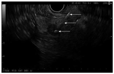
Figure 1 Endoscopic ultrasound-guided fine needle aspiration of a mass in the pancreatic head.
Arrows show the needle course to the tip of the needle within the hypoechoic mass (bottommost arrow).
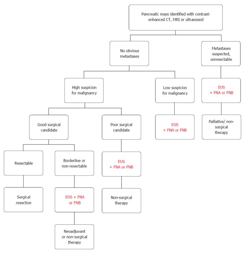
Figure 2 Algorithmic approach to a pancreatic mass.
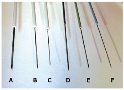
Figure 3 Representative endoscopic ultrasound biopsy and fine needle aspiration needles.
A: 19G core biopsy needle; B: 22G core biopsy needle; C: 25G core biopsy needle; D: 19G FNA needle; E: 22G FNA needle; F: 25G FNA needle.
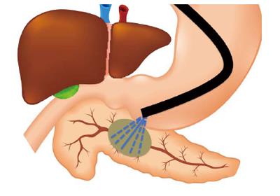
Figure 4 Schematization of the “fanning” technique for endoscopic ultrasound-guided fine needle aspiration.
Dashed lines represent the change in course of the aspiration needle during each needle pass.
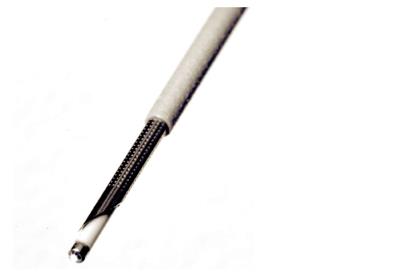
Figure 5 Confocal laser endomicroscopy miniprobe through a 19G FNA needle.
Photo provided with permissions by Mauna Kea, Paris, France.
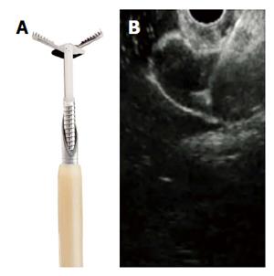
Figure 6 Miniature biopsy forceps.
A: In open position, passing through a 19G FNA needle; B: EUS view of open biopsy forceps through the FNA needle. Photo provided with permissions by US Endoscopy, Mentor, OH.
- Citation: Storm AC, Lee LS. Endoscopic ultrasound-guided techniques for diagnosing pancreatic mass lesions: Can we do better? World J Gastroenterol 2016; 22(39): 8658-8669
- URL: https://www.wjgnet.com/1007-9327/full/v22/i39/8658.htm
- DOI: https://dx.doi.org/10.3748/wjg.v22.i39.8658


