Copyright
©The Author(s) 2015.
World J Gastroenterol. Feb 14, 2015; 21(6): 1827-1837
Published online Feb 14, 2015. doi: 10.3748/wjg.v21.i6.1827
Published online Feb 14, 2015. doi: 10.3748/wjg.v21.i6.1827
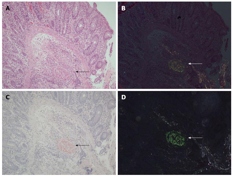
Figure 1 Easily identified amyloid deposits in a submucosal vessel wall (arrows).
A: Brightfield view of hematoxylin-eosin (HE) staining (amyloid is stained pinkish-red); B: Digitally reinforced polarization view of the same HE-stained section. Note the yellowish green tint of birefringence; C: Brightfield view of Congo-red staining (deposits exhibit a salmon-pink appearance); D: Polarization view of the same Congo-red-stained section. Note the apple green tint of polarization is more pronounced than in the digitally reinforced HE polarization image (arrow); 10× magnification.
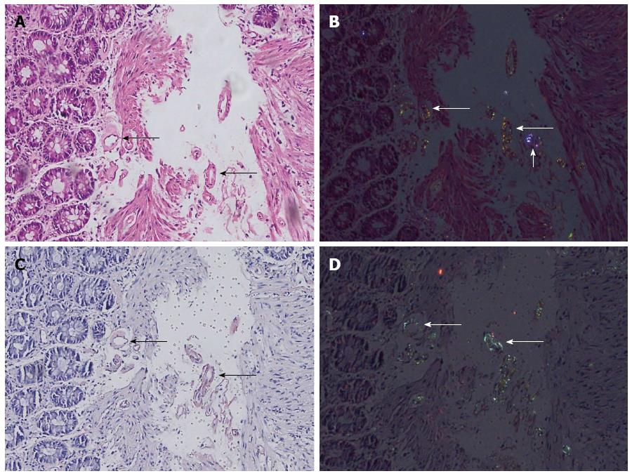
Figure 2 Tiny amyloid deposits in vessel walls, which are not striking at first sight (long arrows).
A: Brightfield view of hematoxylin-eosin (HE) staining; B: Digitally reinforced polarization view of the same HE-stained section. Note the striking yellowish green tint of birefringence of amyloid in small vessel walls (long arrows) and variant colored nonspecific refraction of a foreign body (short arrow); C: Brightfield view of Congo-red staining (note the salmon pink color); D: Polarization view of the same Congo-red-stained section with apple green birefringence of deposits (arrows); 20× magnification.
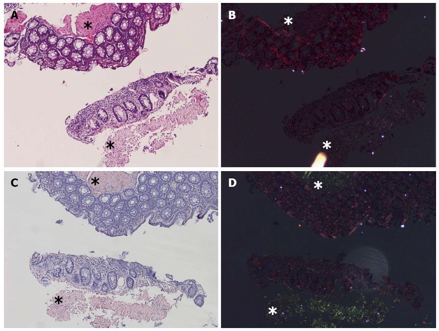
Figure 3 Amyloid suspicious deposits in muscularis mucosa and submucosa (stars).
A: Brightfield view of hematoxylin-eosin (HE) staining; B: Digitally reinforced polarization view of the same HE-stained section. Note the nonspecific refractions throughout the tissue and the aberrant artificial refraction of a foreign body on the slide just below the (stars) in A and B. No convincing birefringence for amyloidosis is observed in the suspected area (stars) by HE; C: Brightfield view of Congo-red staining with salmon pink deposits (stars); D: Polarization view of the same Congo-red-stained section with apple green birefringence of deposits (stars); 10× magnification.
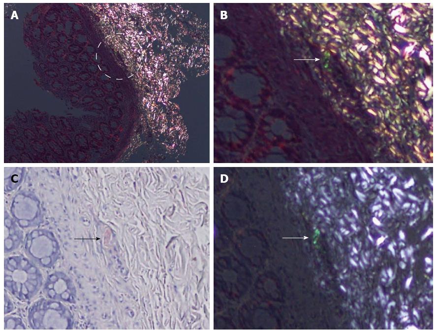
Figure 4 False negative case with extensive refraction and tiny deposits in digitally reinforced polarization view.
A: No arresting positivity was observed in initial evaluation. Note the black-appearing area in the center of the circle [hematoxylin-eosin (HE), under polarization, 10×]; B: High-power digitally reinforced polarization view of the area in circle in panel A. The yellowish green birefringence in a small vessel (arrow) is distinguished after careful examination in comparison with the Congo-red image (HE, under polarization, 40×); C: Congo-red stain positivity in the same vessel (arrow) (bright field, 40×); D: Apple green birefringence of Congo-red polarization of the same vessel (arrow) (under polarization, 40×).
- Citation: Doganavsargil B, Buberal GE, Toz H, Sarsik B, Pehlivanoglu B, Sezak M, Sen S. Digitally reinforced hematoxylin-eosin polarization technique in diagnosis of rectal amyloidosis. World J Gastroenterol 2015; 21(6): 1827-1837
- URL: https://www.wjgnet.com/1007-9327/full/v21/i6/1827.htm
- DOI: https://dx.doi.org/10.3748/wjg.v21.i6.1827


