Copyright
©The Author(s) 2015.
World J Gastroenterol. Jul 7, 2015; 21(25): 7921-7928
Published online Jul 7, 2015. doi: 10.3748/wjg.v21.i25.7921
Published online Jul 7, 2015. doi: 10.3748/wjg.v21.i25.7921
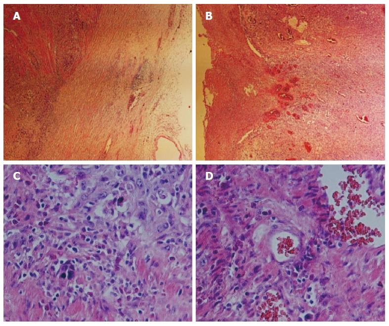
Figure 1 Histopathologic characterization of the esophageal squamous cell carcinoma.
Hematoxylin and eosin staining of the patient’s pathological tissue slide. A and B: Histologic sections revealed a papillary architecture (magnification × 40); C and D: Higher-magnification views of the slides. The tissue presents structural disorder involving abnormal organization, heterotypic cell number, deep nuclear staining, loss of normal epithelial polar structure, and increased mitotic activity. Obvious tumor nests are shown in (D) (magnification × 200).
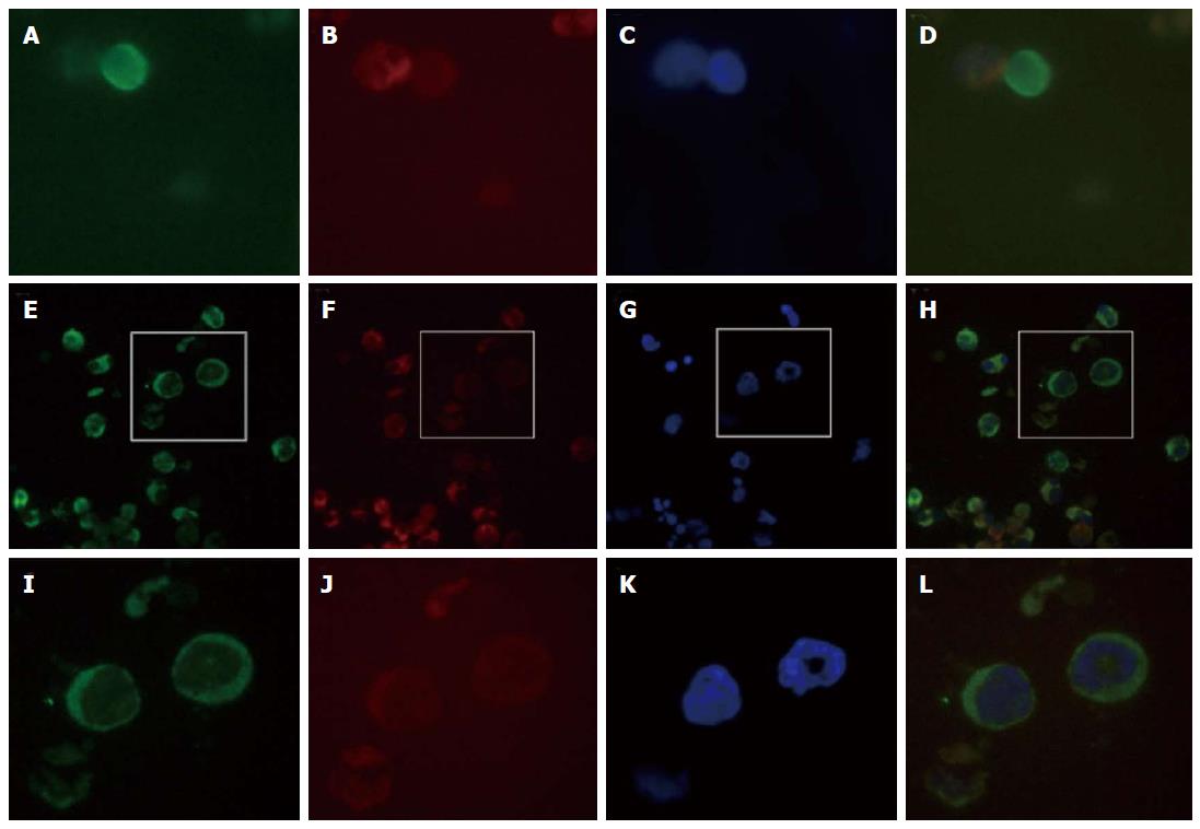
Figure 2 Immunofluorescence analysis of peripheral blood circulating tumor cells collected three months postoperation.
The circulating tumor cells (CTCs) were nucleated and elliptical or elongated, larger than 10 μm, expressed cytokeratin (CK) (CK8/18/19-fluorescein isothiocyanate staining; the epithelial-derived cells are stained in green), lacked CD45 (CD45-phycoerythrin-stained leukocytes are in red), and were positive for 4’,6-diamidino-2-phenylindole (DAPI) nuclear staining (blue-stained nuclei). Some CTCs exhibited morphologically apoptotic features. A single CTC (green) and leukocytes (red) can be observed in fields A-D. A: A CTC stained with anti-CK8/18-FITC (green); B: A leukocyte stained with anti-CD45-PE (red); C: DAPI-stained nuclei; D: Merged image of A-C; E-H: Clusters of tumor cells, CTCs are green (E), leukocytes are red (F), and nuclei are stained with DAPI (G), the merged image of E-G (H); I-K: Enlargements of the boxes above, in which nuclear apoptotic features can be observed in the CTCs.
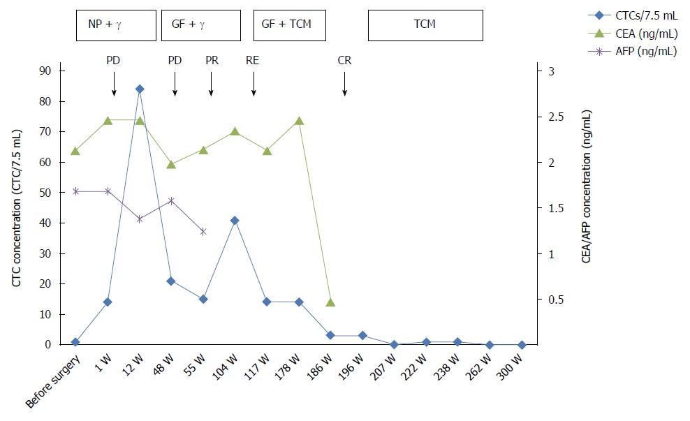
Figure 3 Integrated schema of therapy, biomarkers, and assessments performed.
The various assessments applied during the treatment in this case study are displayed. Boxes representing the duration of treatment are presented at the top of the graph. The names of the therapeutic regimens are shown in the boxes. Changes in circulating tumor cell (CTC) numbers occurred during the different treatments. The carcinoembryonic antigen (CEA) and α-fetoprotein (AFP) markers maintained low or normal values. NP: Cisplatin + vinorelbine; GF: Gemcitabine + leucovorin calcium + fluorouracil; TCM: Traditional Chinese medicine; PD: Progressive disease; PR: Partial response; CR: Complete response; RE: Recurrence.
- Citation: Qiao YY, Lin KX, Zhang Z, Zhang DJ, Shi CH, Xiong M, Qu XH, Zhao XH. Monitoring disease progression and treatment efficacy with circulating tumor cells in esophageal squamous cell carcinoma: A case report. World J Gastroenterol 2015; 21(25): 7921-7928
- URL: https://www.wjgnet.com/1007-9327/full/v21/i25/7921.htm
- DOI: https://dx.doi.org/10.3748/wjg.v21.i25.7921


