Copyright
©The Author(s) 2015.
World J Gastroenterol. Jun 21, 2015; 21(23): 7289-7296
Published online Jun 21, 2015. doi: 10.3748/wjg.v21.i23.7289
Published online Jun 21, 2015. doi: 10.3748/wjg.v21.i23.7289
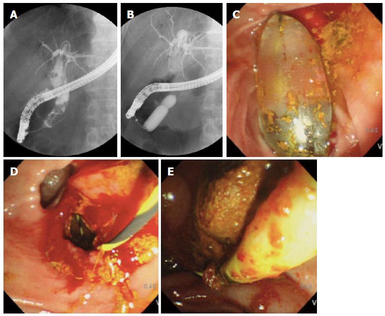
Figure 1 Fluoroscopic and endoscopic view of a bile duct.
A: Multiple large bile duct stones and marked dilation of the common bile duct; B: Endoscopic papillary dilatation with a large (15-18 mm) balloon. Endoscopic sphincterotomy was not performed before balloon sphincteroplasty. This case features incomplete disappearance of the waist; C: An inflated balloon; D: A large biliary orifice was obtained; E: Large stones extraction using a retrieval balloon.
- Citation: Omuta S, Maetani I, Saito M, Shigoka H, Gon K, Tokuhisa J, Naruki M. Is endoscopic papillary large balloon dilatation without endoscopic sphincterotomy effective? World J Gastroenterol 2015; 21(23): 7289-7296
- URL: https://www.wjgnet.com/1007-9327/full/v21/i23/7289.htm
- DOI: https://dx.doi.org/10.3748/wjg.v21.i23.7289


