Copyright
©The Author(s) 2015.
World J Gastroenterol. Mar 28, 2015; 21(12): 3579-3586
Published online Mar 28, 2015. doi: 10.3748/wjg.v21.i12.3579
Published online Mar 28, 2015. doi: 10.3748/wjg.v21.i12.3579
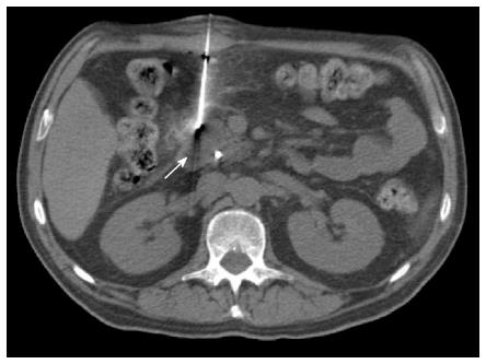
Figure 1 Percutaneous computed tomography-guided core needle biopsy of a pancreatic lesion using direct access.
Non-enhanced axial computed tomography of the upper abdomen showing an anteriorly inserted coaxial needle (17 G) that is placed directly over the lesion located at the head of the pancreas (arrow).
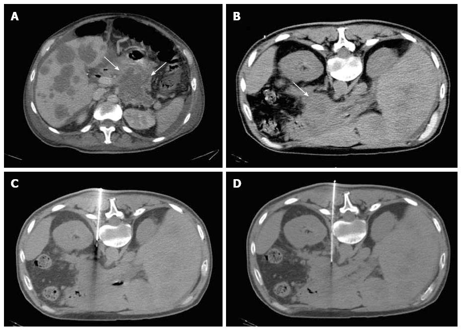
Figure 2 Percutaneous computed tomography-guided core needle biopsy of a pancreatic lesion using hydrodissection.
A: Contrast-enhanced computed tomography (CT) showing an expansive lesion in the tail of the pancreas (arrows). An anterior approach was considered difficult because of the interposition of the intestine in the supine position, therefore, a posterior approach was used; B: Non-enhanced axial CT of the upper abdomen in the prone position showing a lesion located in the tail of the pancreas (arrow); C: Coaxial needle (17 G) positioned in the pillar of the diaphragm, where saline solution was injected to enlarge the paravertebral space, enabling an unobstructed needle path; D: Biopsy needle (18 G) placed adjacent to the pancreatic lesion.
Figure 3 Percutaneous computed tomography-guided core needle biopsy of a pancreatic lesion using pneumodissection.
A: Contrast-enhanced magnetic resonance axial image showing a heterogeneous lesion in the body of the pancreas (arrow). The anterior approach was considered difficult because of the interposition of the intestine in the supine position, therefore, a posterior approach was used; B: Non-enhanced axial computed tomography in the prone position showing the pancreatic lesion (arrow) and the coaxial needle (17 G) positioned in the left pararenal space, where air was injected to displace the kidney and adjacent vessels; C: Biopsy needle (18 G) placed adjacent to the pancreatic lesion.
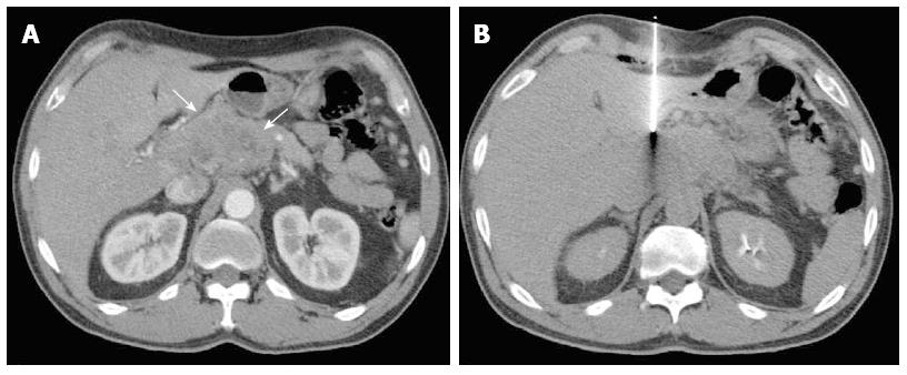
Figure 4 Percutaneous computed tomography-guided core needle biopsy of a pancreatic lesion using transhepatic access.
A: Contrast-enhanced computed tomography showing an expansive lesion in the head and body of the pancreas (arrows). As the patient could not stay in the prone position, the posterior approach was not possible. Therefore, an anterior transhepatic approach was used; B: Biopsy needle (18 G) tip placed adjacent to the pancreatic lesion through the left liver lobe.
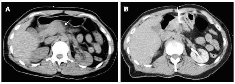
Figure 5 Percutaneous computed tomography-guided core needle biopsy of a pancreatic lesion using transgastric access.
A: Non-enhanced computed tomography showing a poorly defined nodule in the body of the pancreas (arrow). A posterior approach was considered difficult because of the interposition of large vessels in the needle path in the prone position, therefore, an anterior transgastric approach was used; B: Biopsy needle (20 G) placed adjacent to the pancreatic lesion through the stomach.
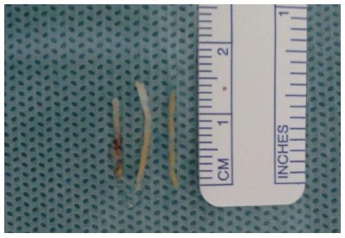
Figure 6 Biopsied specimens of a percutaneous computed tomography-guided core-needle biopsy of a pancreatic lesion.
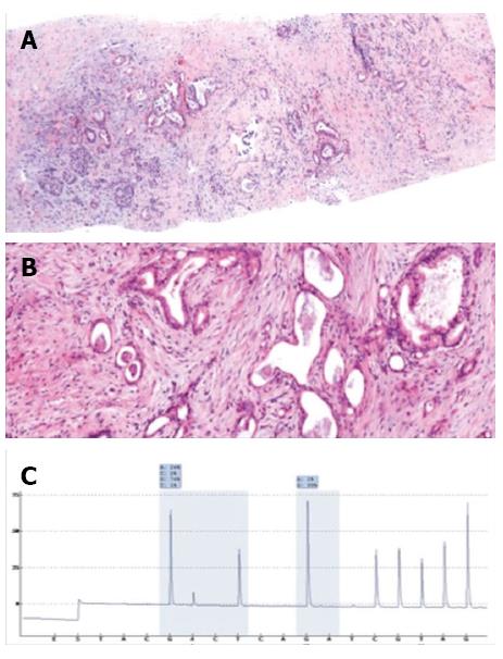
Figure 7 Pathologic and molecular analysis of a biopsied specimen.
A: Panoramic view (× 10) and B: High-power field (× 40) of a pancreatic core biopsy with hematoxylin and eosin staining showing atypical and irregularly displayed ductal structures in a desmoplastic stroma compatible with the diagnosis of ductal adenocarcinoma of the pancreas; C: Pyrogram demonstrating a mutation in codon 12.
- Citation: Tyng CJ, Almeida MFA, Barbosa PN, Bitencourt AG, Berg JAA, Maciel MS, Coimbra FJ, Schiavon LHO, Begnami MD, Guimarães MD, Zurstrassen CE, Chojniak R. Computed tomography-guided percutaneous core needle biopsy in pancreatic tumor diagnosis. World J Gastroenterol 2015; 21(12): 3579-3586
- URL: https://www.wjgnet.com/1007-9327/full/v21/i12/3579.htm
- DOI: https://dx.doi.org/10.3748/wjg.v21.i12.3579


