Copyright
©2014 Baishideng Publishing Group Co.
World J Gastroenterol. Jan 28, 2014; 20(4): 1119-1122
Published online Jan 28, 2014. doi: 10.3748/wjg.v20.i4.1119
Published online Jan 28, 2014. doi: 10.3748/wjg.v20.i4.1119
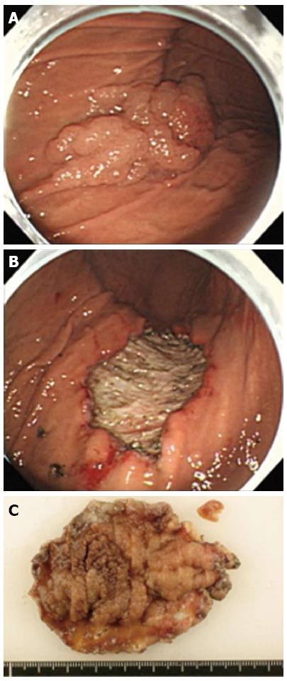
Figure 1 Endoscopic submucosal dissection for early gastric cancer.
A: A cancerous lesion, an early gastric cancer, macroscopic type 0-I+IIa, 75 mm in diameter, is observed on the greater curvature of the gastric body; B: The lesion was successfully removed en bloc without gastric perforation; C: The lesion with the surrounding mucosa was cut into 2-mm-wide serial-step sections.
Figure 2 Abdominal computed tomography and magnetic resonance imaging findings.
A: Abdominal computed tomography (CT) revealed a portion of the thickened gastric wall and a small amount of ascites and free air around the stomach. Free air is indicated by the arrow; B: Abdominal T2-weighted magnetic resonance imaging showed a moderately high-intensity tumor with niveau formation, 5 cm in size, located in the posterior wall of the fundus of the stomach, which suggested focal abscess formation. The abscess is indicated by the arrowhead.
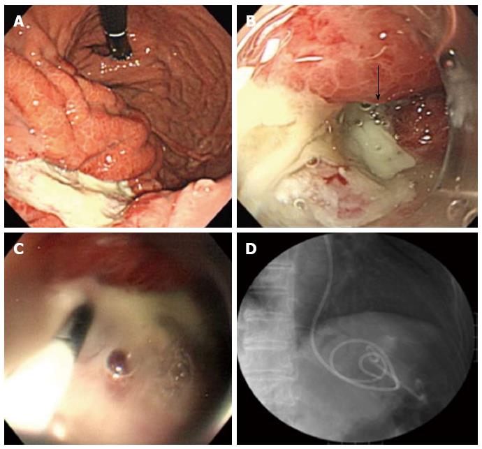
Figure 3 Endoscopic transgastric drainage for a gastric wall abscess.
A: Gastroscopy revealed a smooth, elevated lesion at the greater curvature of the upper body on the oral side of the post-endoscopic submucosal dissection ulcer; B: A gastric fistula was found on the edge of the post-endoscopic submucosal dissection ulcer. The fistula is indicated by an arrow; C: An endoscopic retrograde cholangiopancreatography-catheter was inserted through the fistula into the abscess; D: A plastic stent placement for the internal fistula and a nasocystic catheter for the external fistula were successfully inserted into the abscess.
- Citation: Dohi O, Dohi M, Inoue K, Gen Y, Jo M, Tokita K. Endoscopic transgastric drainage of a gastric wall abscess after endoscopic submucosal dissection. World J Gastroenterol 2014; 20(4): 1119-1122
- URL: https://www.wjgnet.com/1007-9327/full/v20/i4/1119.htm
- DOI: https://dx.doi.org/10.3748/wjg.v20.i4.1119


