Copyright
©2014 Baishideng Publishing Group Inc.
World J Gastroenterol. Aug 14, 2014; 20(30): 10642-10650
Published online Aug 14, 2014. doi: 10.3748/wjg.v20.i30.10642
Published online Aug 14, 2014. doi: 10.3748/wjg.v20.i30.10642
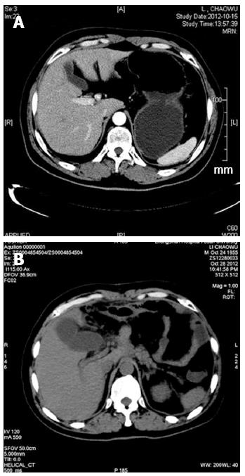
Figure 1 Computed tomography scan in case 1 showing normal gallbladder (A) before operation and a swelling gallbladder with thickened wall (B).
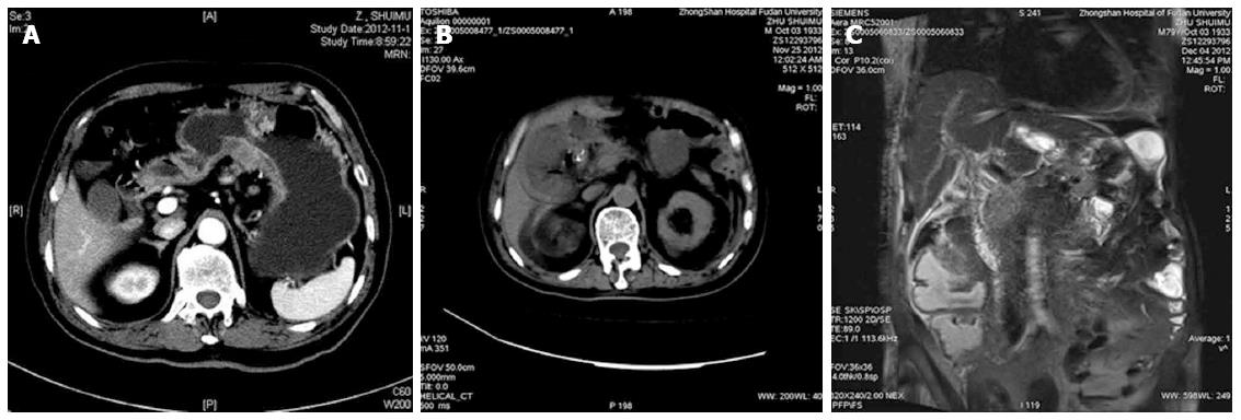
Figure 2 Computed tomography scan in case 2 displaying normal gallbladder (A) before operation, thickened gallbladder wall and swelling gallbladder (B), and magnetic resonance imaging showing no dilation of bile ducts on the 19th day after operation (C).
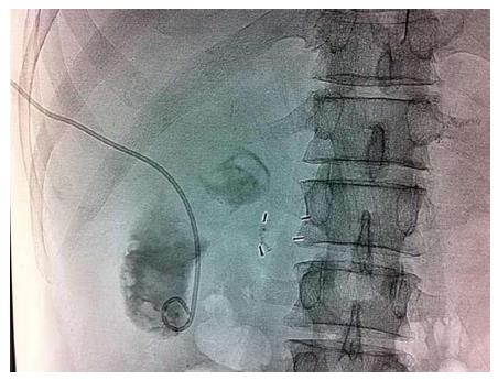
Figure 3 Ultrasound guided percutaneous transhepatic gallbladder drainage in case 3.
- Citation: Liu FL, Li H, Wang XF, Shen KT, Shen ZB, Sun YH, Qin XY. Acute acalculous cholecystitis immediately after gastric operation: Case report and literatures review. World J Gastroenterol 2014; 20(30): 10642-10650
- URL: https://www.wjgnet.com/1007-9327/full/v20/i30/10642.htm
- DOI: https://dx.doi.org/10.3748/wjg.v20.i30.10642


