Copyright
©2013 Baishideng Publishing Group Co.
World J Gastroenterol. Dec 14, 2013; 19(46): 8515-8526
Published online Dec 14, 2013. doi: 10.3748/wjg.v19.i46.8515
Published online Dec 14, 2013. doi: 10.3748/wjg.v19.i46.8515
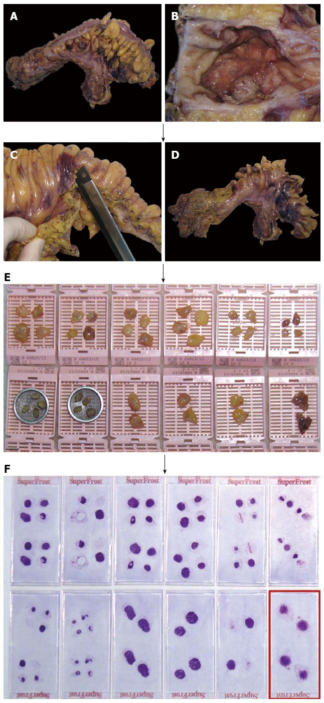
Figure 1 Manual dissection with subsequent histological assessment based on routinely hematoxylin and eosin stained slides is the standard approach in the examination of regional lymph nodes in cancer specimens.
A: Rectum cancer specimen of a 56-year-old female; B: Ulcerated primary tumor, measuring 5 cm in largest diameter; C: After preparation of the primary tumor (including the fatty tissue underneath the lesion and the circumferential margin) the remaining perirectal/mesocolic fatty tissue is carefully removed; D: Specimen for subsequent manual lymph node dissection; E: 36 presumed lymph nodes are isolated, of which the largest four are cut into halves and embedded on their own, respectively (lower right); F: 31 lymph nodes are confirmed on hematoxylin and eosin stained slides, one of which with metastatic cancer tissue (encircled).
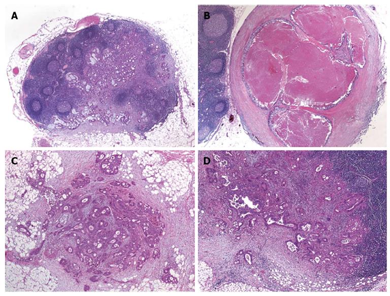
Figure 2 Lymph node metastases and tumor deposits in patients with colorectal cancer.
A: Metastatic adenocarcinoma within a mesocolic lymph node [hematoxylin and eosin (HE) original magnification, × 100]; B: Mesocolic lymph node totally replaced by metastatic cancer tissue, note the smooth contour of the lesion (HE, original magnification, × 150); C: Tumor deposit (satellite) within the mesocolic fatty tissue, note the irregular contour of the lesion (HE, original magnification, × 250); D: Mesocolic lymph node metastasis with extracapsular extension of cancer tissue (original magnification, × 250).
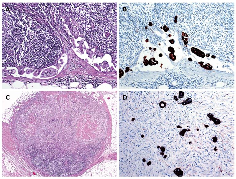
Figure 3 Value of immunohistochemistry in the evaluation of lymph nodes from patients with colorectal cancer.
A: Micrometastasis in the subcapsular sinus of a mesocolic sentinel node evaluated by standard hematoxylin and eosin (HE) staining (original magnification, × 400); B: Micrometastasis in the subcapsular sinus of a mesocolic sentinel node evaluated by immunohistochemistry using an antibody preparation directed against pankeratin (serial section to A, original magnification, × 400); C: Atrophic perirectal lymph node with marked fibrosis after neoadjuvant treatment (original magnification, × 100); D: Identification of residual cancer cells by pankeratin immunostaining (original magnification, × 400).
- Citation: Resch A, Langner C. Lymph node staging in colorectal cancer: Old controversies and recent advances. World J Gastroenterol 2013; 19(46): 8515-8526
- URL: https://www.wjgnet.com/1007-9327/full/v19/i46/8515.htm
- DOI: https://dx.doi.org/10.3748/wjg.v19.i46.8515


