Copyright
©2013 Baishideng Publishing Group Co.
World J Gastroenterol. Oct 14, 2013; 19(38): 6353-6359
Published online Oct 14, 2013. doi: 10.3748/wjg.v19.i38.6353
Published online Oct 14, 2013. doi: 10.3748/wjg.v19.i38.6353
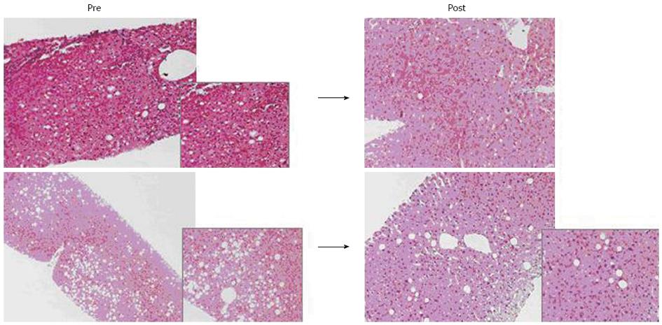
Figure 1 Two histologic examinations of liver parenchyma with steatosis, before and after intensive dietary treatment in the same donor.
10/18 donors successfully completed the 3-mo treatment (body mass index < 30 kg/m²). In all 10 treated donors, hepatic macrosteatosis < 30% was found (range: 2%-15%) and therefore they were considered eligible for donation.
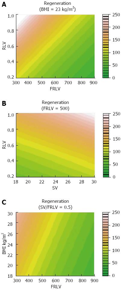
Figure 2 Locally Weighted Scatterplot Smoother graphics of the overall population, evidencing the association of percentage of liver regeneration with levels of body mass index (A), future remnant liver volume (B) and spleen volume/future remnant liver volume (C).
FRLV: Future remnant liver volume; SV: Spleen volume; BMI: Body mass index.
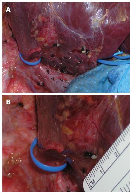
Figure 3 A large accessory hepatic vein draining the right lobe along with the right hepatic vein is encircled using a vessel loop (A).
The measurement of the accessory hepatic vein [major of 5 mm, (B)] was performed for decision making to transect it with a vascular stapler, only when transections of liver parenchyma and of the structures of hepatic pedicle were almost complete.
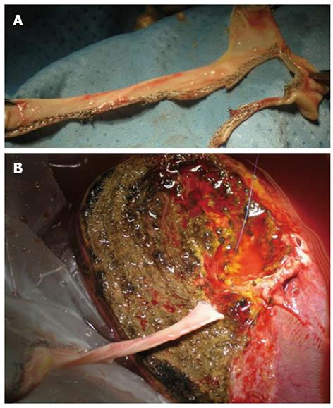
Figure 4 Usage of several vascular staplers were crucial to shapes and employees a heterologous venous conduit (A), previously harvested from a deceased donors, for a back-table reconstruction of a large accessory hepatic vein (B) to be connected to the recipient inferior vena cava in the LERLT.
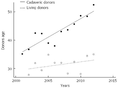
Figure 5 Linear regression models were used for explaining differences in terms of age between deceased and living liver donors over the last decade at the Mediterranean Institute for Transplantation and Advanced Specialized Therapies, and the University of Pittsburgh Medical Center in Italy.
In our series of 677 cadaveric liver transplantation and 105 living donor liver transplantation, the deceased donor age has been increasing with a significative difference of average values between deceased and living liver donor age (95%CI: 0.9-1.3), P < 0.0001.
- Citation: Gruttadauria S, Pagano D, Cintorino D, Arcadipane A, Traina M, Volpes R, Luca A, Vizzini G, Gridelli B, Spada M. Right hepatic lobe living donation: A 12 years single Italian center experience. World J Gastroenterol 2013; 19(38): 6353-6359
- URL: https://www.wjgnet.com/1007-9327/full/v19/i38/6353.htm
- DOI: https://dx.doi.org/10.3748/wjg.v19.i38.6353


