Copyright
©2013 Baishideng Publishing Group Co.
World J Gastroenterol. Aug 21, 2013; 19(31): 5035-5050
Published online Aug 21, 2013. doi: 10.3748/wjg.v19.i31.5035
Published online Aug 21, 2013. doi: 10.3748/wjg.v19.i31.5035
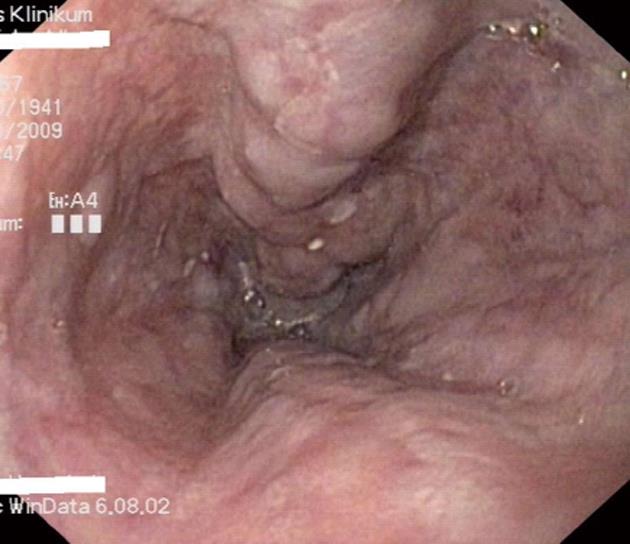
Figure 1 Esophageal varices grade II in a patient with liver cirrhosis.
Figure 2 Variceal band ligation of esophageal varices.
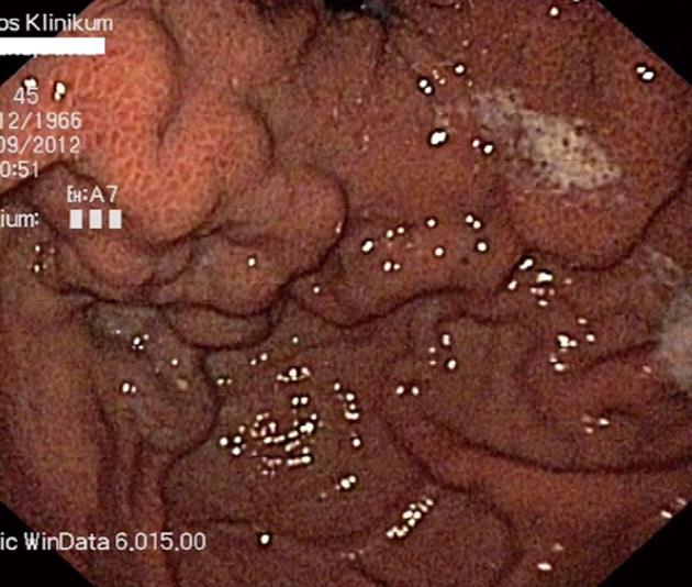
Figure 3 Isolated gastric varices type I and portal hypertensive gastropathy in a patient with liver cirrhosis.
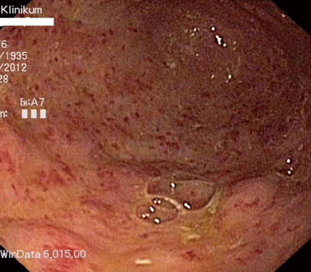
Figure 4 Acute diffuse bleeding from portal hypertensive gastropathy in a patient with decompensated liver cirrhosis.
Figure 5 Typical appearance of a watermelon stomach in a patient with gastric antral vascular ectasia-syndrome and compensated liver cirrhosis.
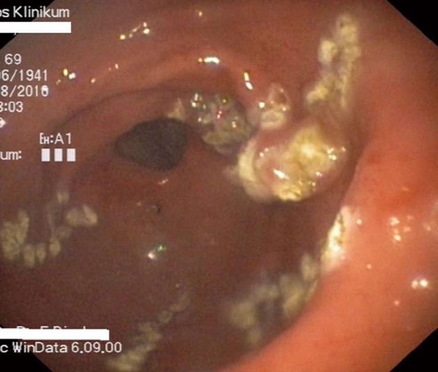
Figure 6 Endoscopic treatment of gastric antral vascular ectasia with argon plasma coagulation therapy.
- Citation: Biecker E. Portal hypertension and gastrointestinal bleeding: Diagnosis, prevention and management. World J Gastroenterol 2013; 19(31): 5035-5050
- URL: https://www.wjgnet.com/1007-9327/full/v19/i31/5035.htm
- DOI: https://dx.doi.org/10.3748/wjg.v19.i31.5035


