Copyright
©2013 Baishideng Publishing Group Co.
World J Gastroenterol. Jan 21, 2013; 19(3): 404-410
Published online Jan 21, 2013. doi: 10.3748/wjg.v19.i3.404
Published online Jan 21, 2013. doi: 10.3748/wjg.v19.i3.404
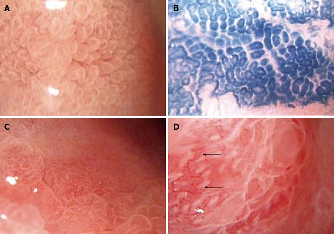
Figure 1 Magnifying chromoendoscopic findings.
A: Type E pits; B: Type E pits after dye staining; C: Type F pits; D: Type F pits after dye staining, arrows show disorderly arranged thickened capillaries. Magnification, ×100. Type E: Villous pits, with finger-like tubers similar to enteral villus-like changes; Type F: The pits have obscure or disappearing structures and extremely irregular arrangement.
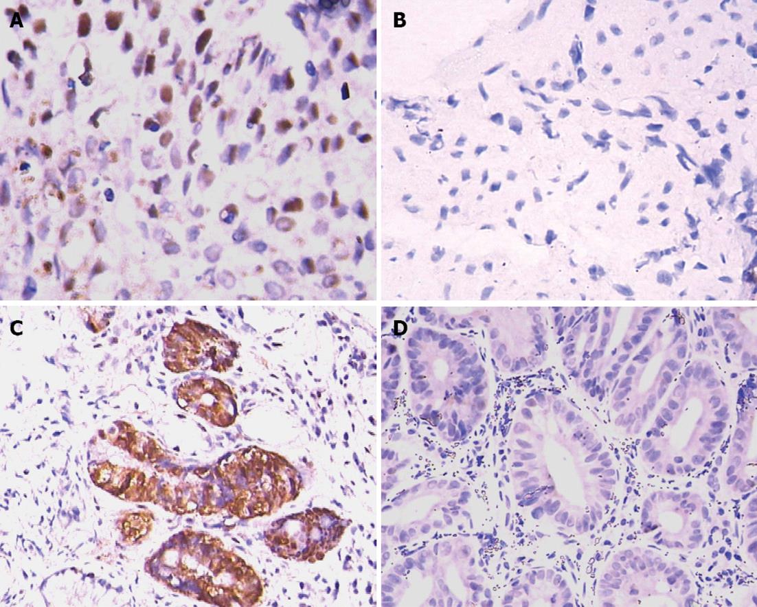
Figure 2 Immunohistochemical results.
A: Expression of p53 in gastric pits in chronic atrophic gastritis, and nuclei were stained dark brown; B: p53 expression was undetectable in gastric pits in chronic superficial gastritis; C: Expression of proliferating cell nuclear antigen (PCNA) was detected in gastric pits in chronic atrophic gastritis, and nuclei were stained dark brown; D: PCNA expression was undetectable in gastric pits in chronic superficial gastritis.
- Citation: Meng XM, Zhou Y, Dang T, Tian XY, Kong J. Magnifying chromoendoscopy combined with immunohistochemical staining for early diagnosis of gastric cancer. World J Gastroenterol 2013; 19(3): 404-410
- URL: https://www.wjgnet.com/1007-9327/full/v19/i3/404.htm
- DOI: https://dx.doi.org/10.3748/wjg.v19.i3.404


