Copyright
©2013 Baishideng Publishing Group Co.
World J Gastroenterol. Apr 21, 2013; 19(15): 2362-2367
Published online Apr 21, 2013. doi: 10.3748/wjg.v19.i15.2362
Published online Apr 21, 2013. doi: 10.3748/wjg.v19.i15.2362
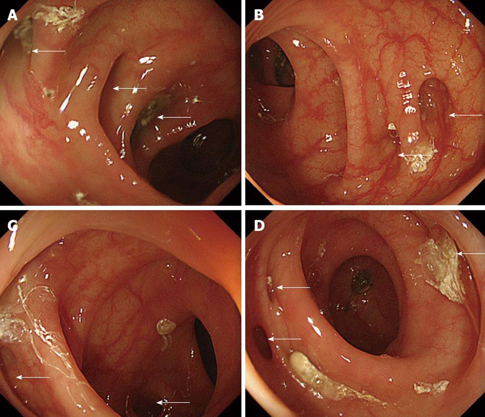
Figure 1 Colonic diverticula in the left colon on endoscopy.
The colon location was classified as the right colon (cecum and ascending and transverse colon) or the left colon (descending colon and sigmoid colon). A: Sigmoid-descending colon junction; B: Proximal sigmoid colon; C: Sigmoid top; D: Distal sigmoid colon. Arrows show colonic diverticula determined by colonoscopy.
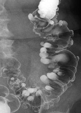
Figure 2 Colonic diverticula in the left colon on radiography with barium enema.
The colonic location was classified as the right colon (cecum and ascending and transverse colon) or the left colon (descending colon and sigmoid colon).
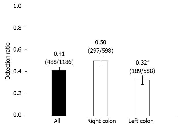
Figure 3 Detection ratio (colonoscopy/barium) of colonic diverticula in 65 patients.
Right colon denotes the cecum, ascending colon, and transverse colon. Left colon denotes the descending colon and sigmoid colon. aP < 0.05 by χ2 test. Error bars show the 95%CI of the detection ratio.
- Citation: Niikura R, Nagata N, Shimbo T, Akiyama J, Uemura N. Colonoscopy can miss diverticula of the left colon identified by barium enema. World J Gastroenterol 2013; 19(15): 2362-2367
- URL: https://www.wjgnet.com/1007-9327/full/v19/i15/2362.htm
- DOI: https://dx.doi.org/10.3748/wjg.v19.i15.2362


