Copyright
©2013 Baishideng Publishing Group Co.
World J Gastroenterol. Mar 14, 2013; 19(10): 1618-1624
Published online Mar 14, 2013. doi: 10.3748/wjg.v19.i10.1618
Published online Mar 14, 2013. doi: 10.3748/wjg.v19.i10.1618
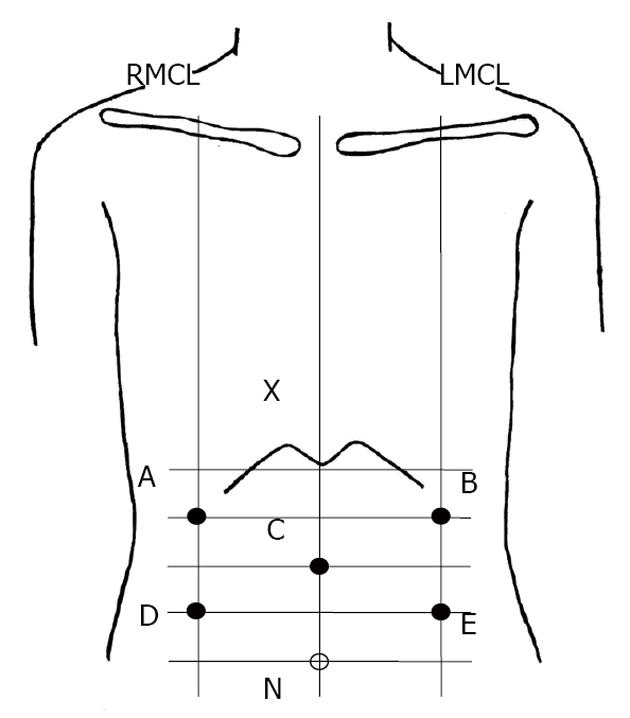
Figure 1 Positions of electrodes for electrogastrography recording.
X: Xyphoid process; N: Navel; A: Channel 1 placed on the intersecting point between the right mid-clavicular line (RMCL) and the vertical bisectrix of the line XC; B: Channel 2 placed on the intersecting point between left mid-clavicular line (LMCL) the avertical bisectrix of the line XC; C: Central terminal electrode placed on the patient’s ventral midline approximately halfway between the umbilicus and the xyphoid process; D: Channel 3 placed on the intersecting point between RMCL and the avertical bisectrix of the line NC; E: Channel 4 placed on the intersecting point between LMCL and a vertical bisectrix of the line NC.
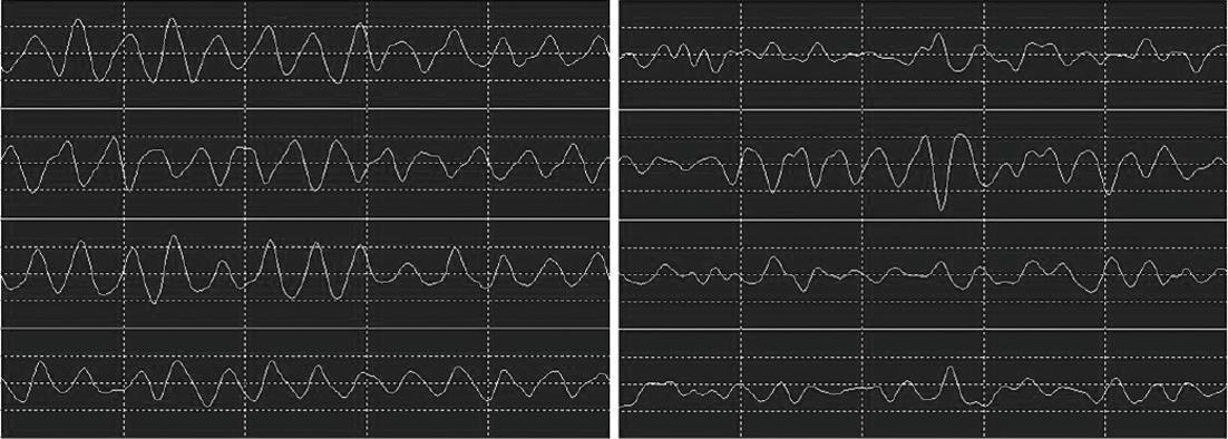
Figure 2 Example of the raw electrogastrography signals before percutaneous local therapy for hepatocellular carcinoma (left) and after therapy (right).
- Citation: Kobayashi M, Kinekawa F, Matsuda K, Fujihara S, Nishiyama N, Nomura T, Tani J, Miyoshi H, Kobara H, Deguchi A, Yoneyama H, Mori H, Masaki T. Influence of percutaneous local therapy for hepatocellular carcinoma on gastric function. World J Gastroenterol 2013; 19(10): 1618-1624
- URL: https://www.wjgnet.com/1007-9327/full/v19/i10/1618.htm
- DOI: https://dx.doi.org/10.3748/wjg.v19.i10.1618


