Copyright
©2012 Baishideng Publishing Group Co.
World J Gastroenterol. Dec 21, 2012; 18(47): 7122-7126
Published online Dec 21, 2012. doi: 10.3748/wjg.v18.i47.7122
Published online Dec 21, 2012. doi: 10.3748/wjg.v18.i47.7122
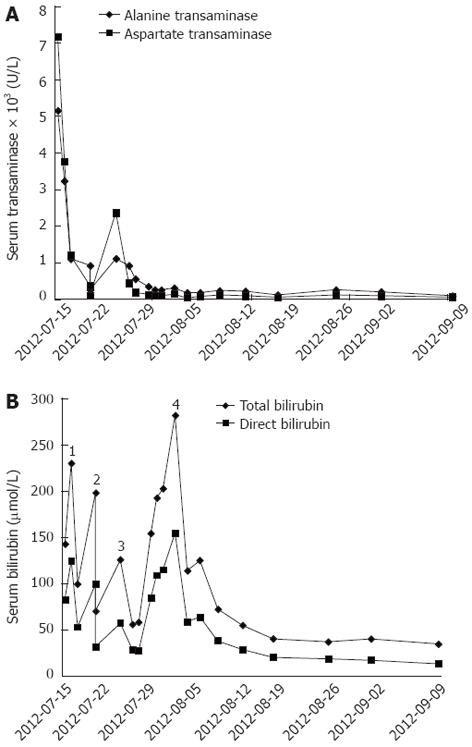
Figure 1 Laboratory test results of physical examination.
A: Blood concentration of transaminases markedly decreased after artificial liver support (ALS)-plasma exchange, increased after liver transplantation due to ischemia reperfusion injury, and reached a peak on post-transplantation day 2, then decreased gradually to normal; B: Blood concentration of bilirubin. There are four marked peaks: bilirubin increased due to fulminant liver failure and decreased after two ALS-plasma exchanges, forming peaks 1 and 2; bilirubin increased after liver transplantation due to ischemia reperfusion injury and reached a peak, forming peak 3; bilirubin increased markedly because of the intracholedochal hematoma and decreased after percutaneous transhepatic intervention, forming peak 4.
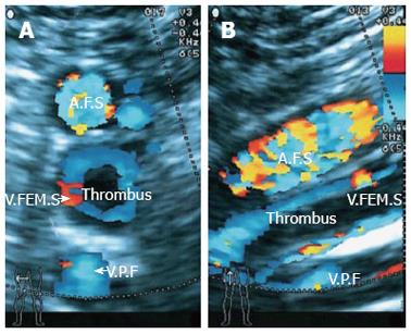
Figure 2 Doppler ultrasound shows a thrombus in the right femoral vein.
A: Axial plane; B: Sagittal plane. A.F.S.: Superficial femoral artery; V.FEM.S: Superficial femoral vein; V.P.F: Profound femoral vein.
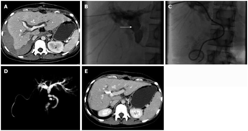
Figure 3 Diagnosis of obstructive jaundice.
A: Before percutaneous transhepatic intervention, on computed tomography (CT) scan, intrahepatic biliary ducts are dilated and a huge hematoma is present below the right lobe of liver; B: Percutaneous transhepatic cholangiography shows a long intrabiliary filling defect (white arrow) crossing the strictured biliary anastomosis (black arrow); C: An 8F biliary drainage catheter was placed across the anastomosis with one end opening into the duodenum and the other end exiting the body; D: On magnetic resonance cholangiopancreatography, the intrahepatic biliary ducts were slightly dilated at the first follow-up; E: CT reveals that the hematoma below the liver was almost absorbed and the intrahepatic biliary ducts were slightly dilated at the first follow-up.
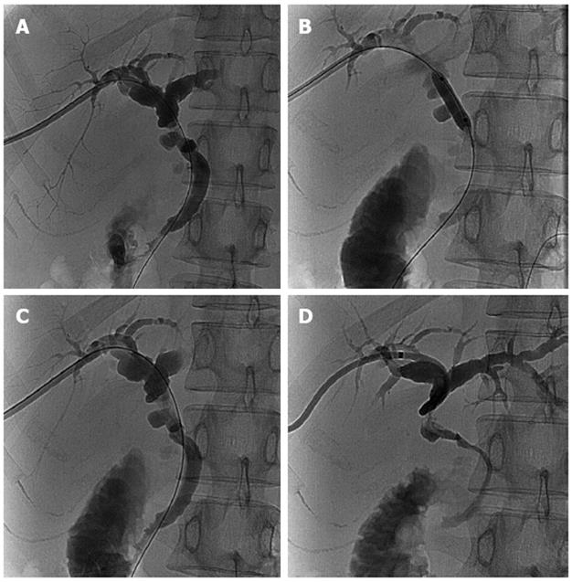
Figure 4 Cholangiography was performed through the biliary drainage catheter.
A: At the first follow-up, percutaneous transhepatic cholangiography shows the slightly dilated intrahepatic biliary ducts and a stricture at the site of anastomosis; B: Balloon dilatation of the anastomotic stricture; C: Anastomotic stricture was relieved after balloon dilation; D: A new biliary drainage catheter was placed.
- Citation: Qin JJ, Xia YX, Lv L, Wang ZJ, Zhang F, Wang XH, Sun BC. Successful disintegration, dissolution and drainage of intracholedochal hematoma by percutaneous transhepatic intervention. World J Gastroenterol 2012; 18(47): 7122-7126
- URL: https://www.wjgnet.com/1007-9327/full/v18/i47/7122.htm
- DOI: https://dx.doi.org/10.3748/wjg.v18.i47.7122


