Copyright
©2012 Baishideng Publishing Group Co.
World J Gastroenterol. Jun 28, 2012; 18(24): 3050-3057
Published online Jun 28, 2012. doi: 10.3748/wjg.v18.i24.3050
Published online Jun 28, 2012. doi: 10.3748/wjg.v18.i24.3050
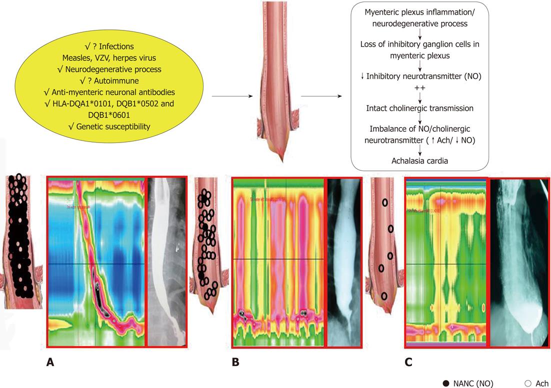
Figure 1 Schematic diagram outlining possible pathogenesis of achalasia cardia.
A: Diagram showing distribution of nitric oxide (NO) and acetycholine (Ach) neurons in the esophagus with normal motility pattern and barium esophagogram; B: Loss of NO in esophagus in early stage of the disease resulting in high amplitude simultaneous contractions in body (called vigorous achalasia). In this stage esophagus is not dilated in barium esophagogram; C: Further degeneration of inhibitory and additional degeneration of excitatory neurons causes low amplitude simultaneous contractions in esophageal body (called classic achalasia). In this stage esophagus is dilated in barium esophagogram. NANC: Non-adrenergic, non-cholinergic.
- Citation: Ghoshal UC, Daschakraborty SB, Singh R. Pathogenesis of achalasia cardia. World J Gastroenterol 2012; 18(24): 3050-3057
- URL: https://www.wjgnet.com/1007-9327/full/v18/i24/3050.htm
- DOI: https://dx.doi.org/10.3748/wjg.v18.i24.3050


