Copyright
©2012 Baishideng Publishing Group Co.
World J Gastroenterol. May 7, 2012; 18(17): 2145-2146
Published online May 7, 2012. doi: 10.3748/wjg.v18.i17.2145
Published online May 7, 2012. doi: 10.3748/wjg.v18.i17.2145
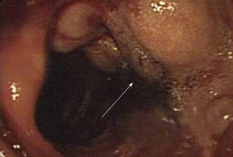
Figure 1 Enteroscopy revealed a mass at the small intestine (arrow).
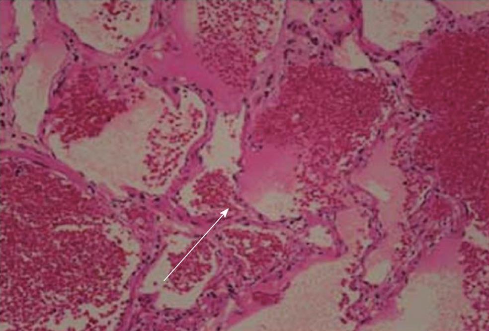
Figure 2 Histological analysis revealed a benign soft tissue mass (arrow) consisting of lymphatic and blood vessels (hematoxylin and eosin, × 100).
- Citation: Fang YF, Qiu LF, Du Y, Jiang ZN, Gao M. Small intestinal hemolymphangioma with bleeding: A case report. World J Gastroenterol 2012; 18(17): 2145-2146
- URL: https://www.wjgnet.com/1007-9327/full/v18/i17/2145.htm
- DOI: https://dx.doi.org/10.3748/wjg.v18.i17.2145


