Copyright
©2012 Baishideng Publishing Group Co.
World J Gastroenterol. Apr 7, 2012; 18(13): 1496-1501
Published online Apr 7, 2012. doi: 10.3748/wjg.v18.i13.1496
Published online Apr 7, 2012. doi: 10.3748/wjg.v18.i13.1496
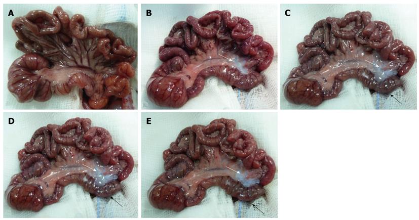
Figure 1 Macroscopic monitoring.
A: Physiological appearance of the rat bowel; B: Rat bowel 1 h after inferior mesenteric artery (IMA) ligation; C: At 4 h after IMA ligation; D: At 6 h after IMA ligation; E: At 8 h after IMA ligation.
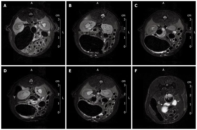
Figure 2 7T magnetic resonance imaging investigation.
A: Image of a 7T magnetic resonance imaging (MRI) abdominal scan before inferior mesenteric artery (IMA) ligation; B: A 7T MRI abdominal scan 1 h after IMA ligation; C: At 4 h after IMA ligation; D: At 6 h after IMA ligation; E: At 8 h after IMA ligation; F: Image of 7T MRI colon enema.
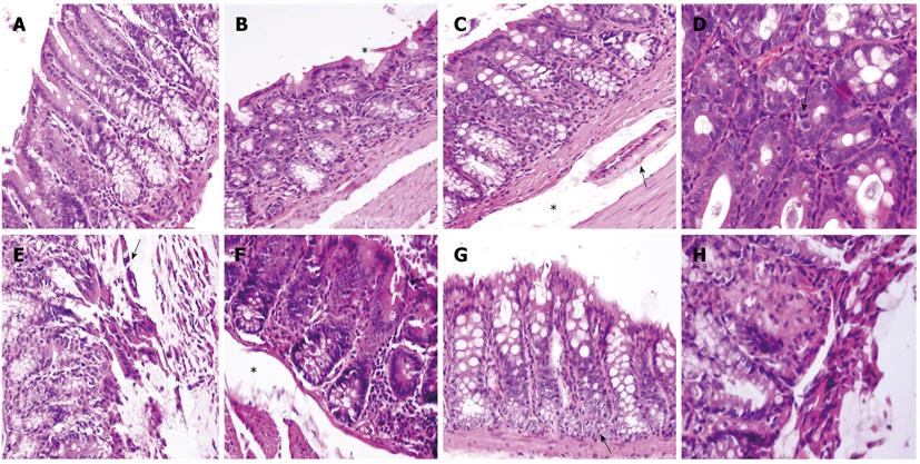
Figure 3 Histological analysis.
A: Normal histological pattern of the rat proximal colon; B: Histological pattern of the rat proximal colon 1 h after inferior mesenteric artery (IMA) ligation; C: At 4 h after IMA ligation; D: At 6 h after IMA ligation; E: At 8 h after IMA ligation; note the necrosis and loss of the surface epithelium; F: Markedly edematous submucosa (star); G: Active regeneration signs at the bottom of the crypts; H: Coagulative necrosis.
- Citation: Iacobellis F, Berritto D, Somma F, Cavaliere C, Corona M, Cozzolino S, Fulciniti F, Cappabianca S, Rotondo A, Grassi R. Magnetic resonance imaging: A new tool for diagnosis of acute ischemic colitis? World J Gastroenterol 2012; 18(13): 1496-1501
- URL: https://www.wjgnet.com/1007-9327/full/v18/i13/1496.htm
- DOI: https://dx.doi.org/10.3748/wjg.v18.i13.1496


