Copyright
©2011 Baishideng Publishing Group Co.
World J Gastroenterol. Dec 14, 2011; 17(46): 5059-5074
Published online Dec 14, 2011. doi: 10.3748/wjg.v17.i46.5059
Published online Dec 14, 2011. doi: 10.3748/wjg.v17.i46.5059
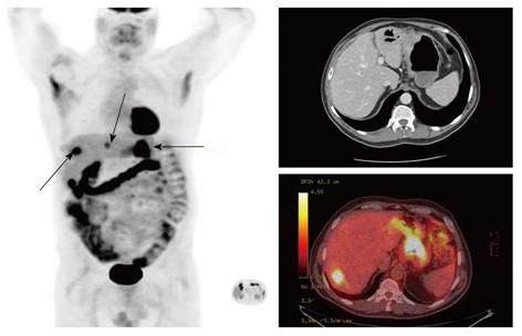
Figure 1 (18F) 2-fluoro-2-deoxyglucose-positron emission tomography/computed tomography image of a patient with a proximal gastric cancer and occult liver metastasis.
The liver lesion was not identified on the corresponding staging computed tomography.
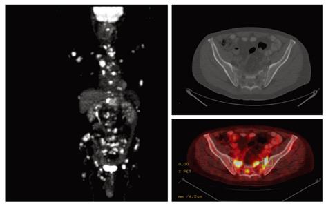
Figure 2 (18F) 2-fluoro-2-deoxyglucose-positron emission tomography/computed tomography detects diffuse bony metastases not seen on staging computed tomography.
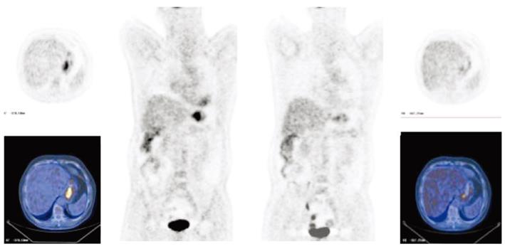
Figure 3 (18F) 2-fluoro-2-deoxyglucose-positron emission tomography response in a patient with a proximal gastric cancer receiving chemotherapy.
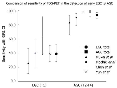
Figure 4 Sensitivity of (18F) 2-fluoro-2-deoxyglucose-positron emission tomography to identify primary early and advanced gastric carcinoma.
FDG-PET: (18F) 2-fluoro-2-deoxyglucose-positron emission tomography; EGC: Early gastric cancer; AGC: Advanced gastric cancer; CI: Confidence interval.
- Citation: Smyth EC, Shah MA. Role of (18F) 2-fluoro-2-deoxyglucose positron emission tomography in upper gastrointestinal malignancies. World J Gastroenterol 2011; 17(46): 5059-5074
- URL: https://www.wjgnet.com/1007-9327/full/v17/i46/5059.htm
- DOI: https://dx.doi.org/10.3748/wjg.v17.i46.5059


