Copyright
©2011 Baishideng Publishing Group Co.
World J Gastroenterol. Aug 14, 2011; 17(30): 3560-3564
Published online Aug 14, 2011. doi: 10.3748/wjg.v17.i30.3560
Published online Aug 14, 2011. doi: 10.3748/wjg.v17.i30.3560
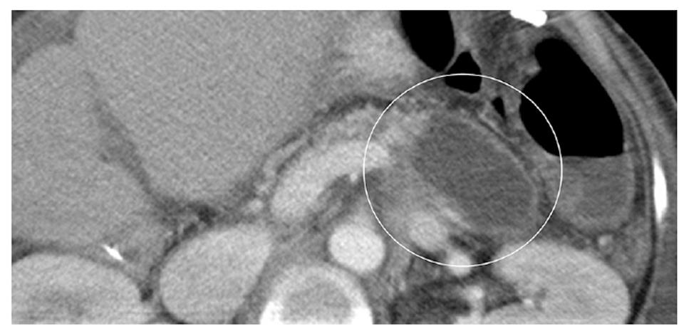
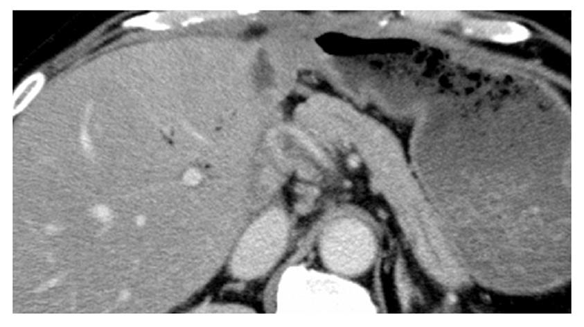
Figure 1 Computed tomography performed on postoperative day 13 showing fluid collection (circle) at the lower edge of the pancreas.
The hematoma was considered to be ruptured.
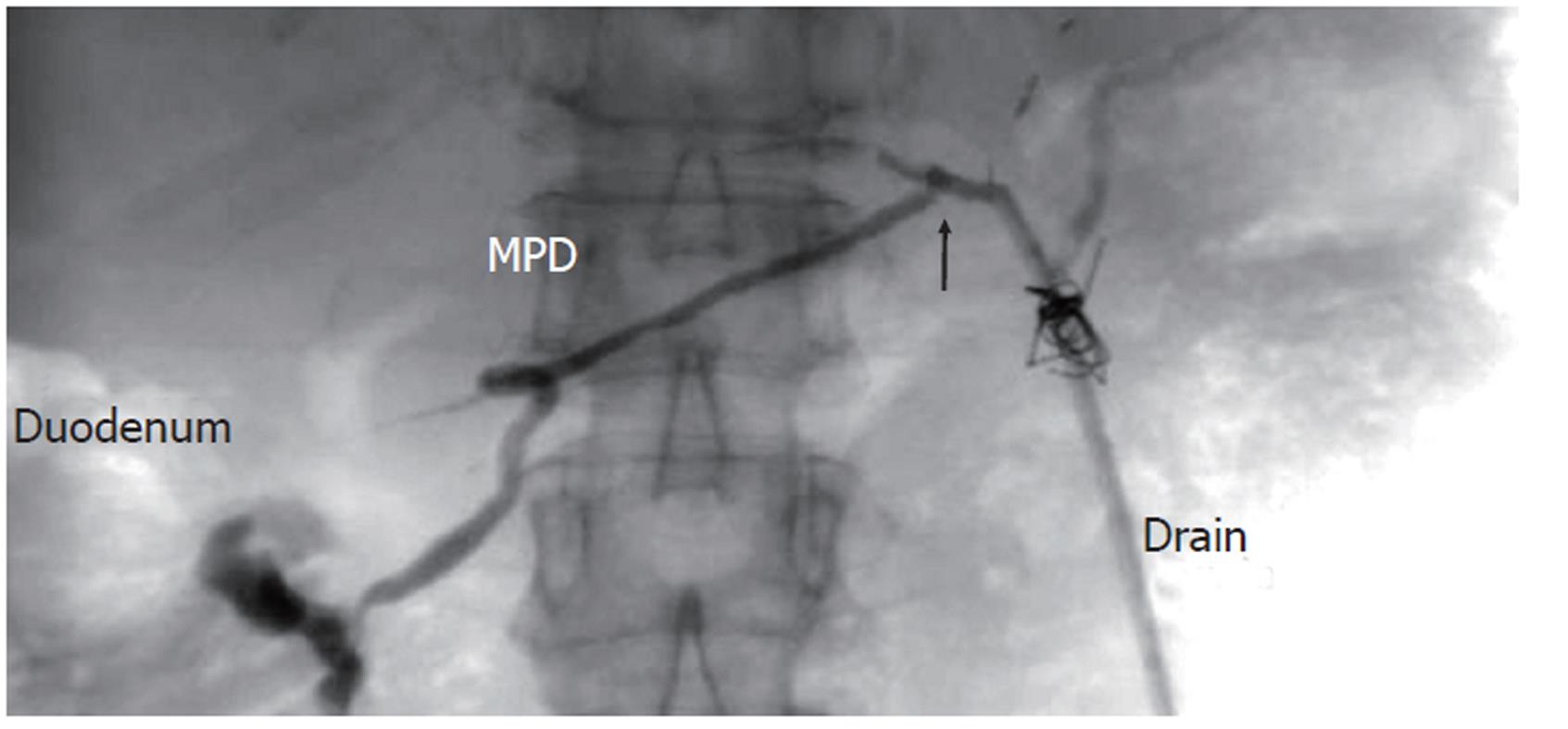
Figure 2 Pancreatographic examination of the drain at the tail of the pancreas (Drain) in case 1 reveals the disrupted (arrow) main pancreatic duct with flow of contrast into the duodenum.
MPD: Main pancreatic duct.
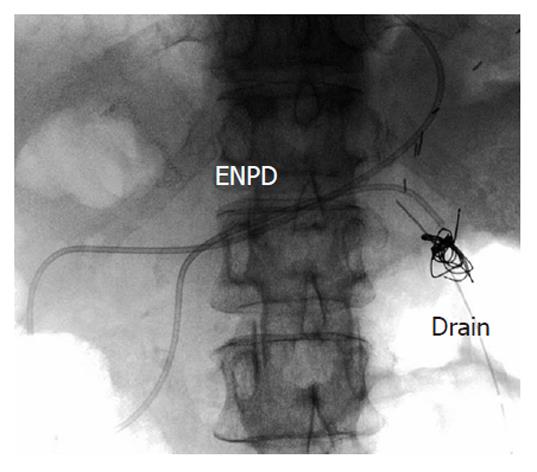
Figure 3 A radiograph showing postprocedure endoscopic naso-pancreatic drainage in case 1.
Excellent drainage of the pancreatic duct is noted. ENPD: Endoscopic naso-pancreatic drainage.
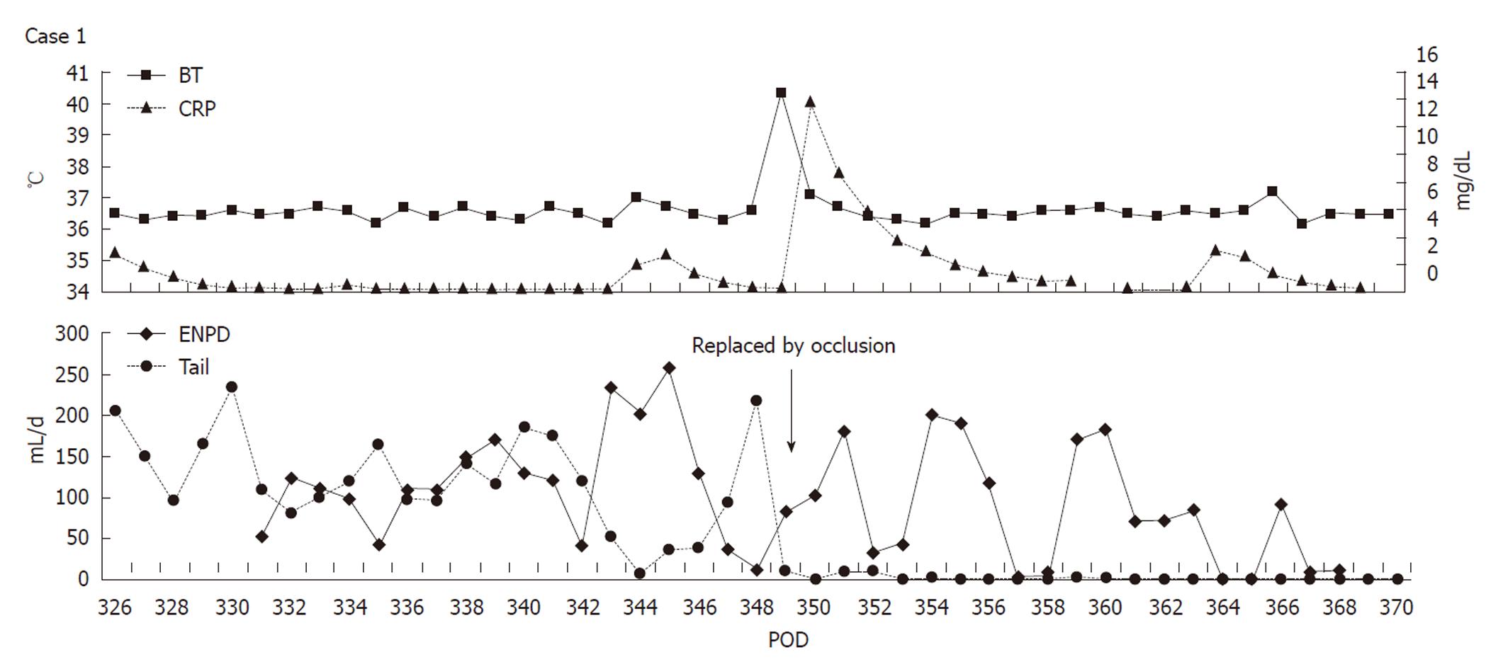
Figure 4 Upper chart shows the body temperature and serum C-reactive protein level.
Lower one shows daily output of the endoscopic naso-pancreatic drainage tube and the drain at the tail of the pancreas (Tail) in case 1. The patient had an episode of fever caused by the occlusion of the endoscopic naso-pancreatic drainage (ENPD) tube. After the tube was replaced, the pancreatic fistula healed completely. BT: Body temperature; CRP: C-reactive protein; POD: Postoperative day.
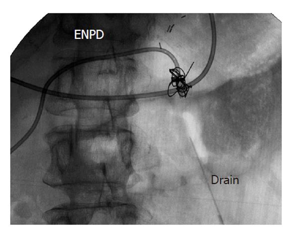
Figure 5 Contrast examination from the drain at the tail of the pancreas (Drain) in case 1 on postoperative day 363 reveals the closure of fistula.
ENPD: Endoscopic naso-pancreatic drainage.
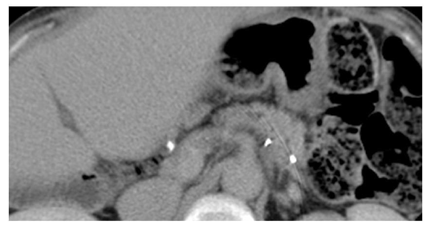
Figure 6 Computed tomography performed 2 d after removal of the drain in case 1 showing no fluid collection around the pancreas.
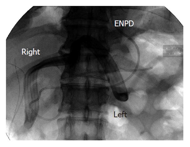
Figure 7 A radiograph showing postprocedure endoscopic naso-pancreatic drainage in case 2.
The image shows drains placed at both the right and left edges of the pancreas (Right and Left) and endoscopic naso-pancreatic drainage (ENPD).
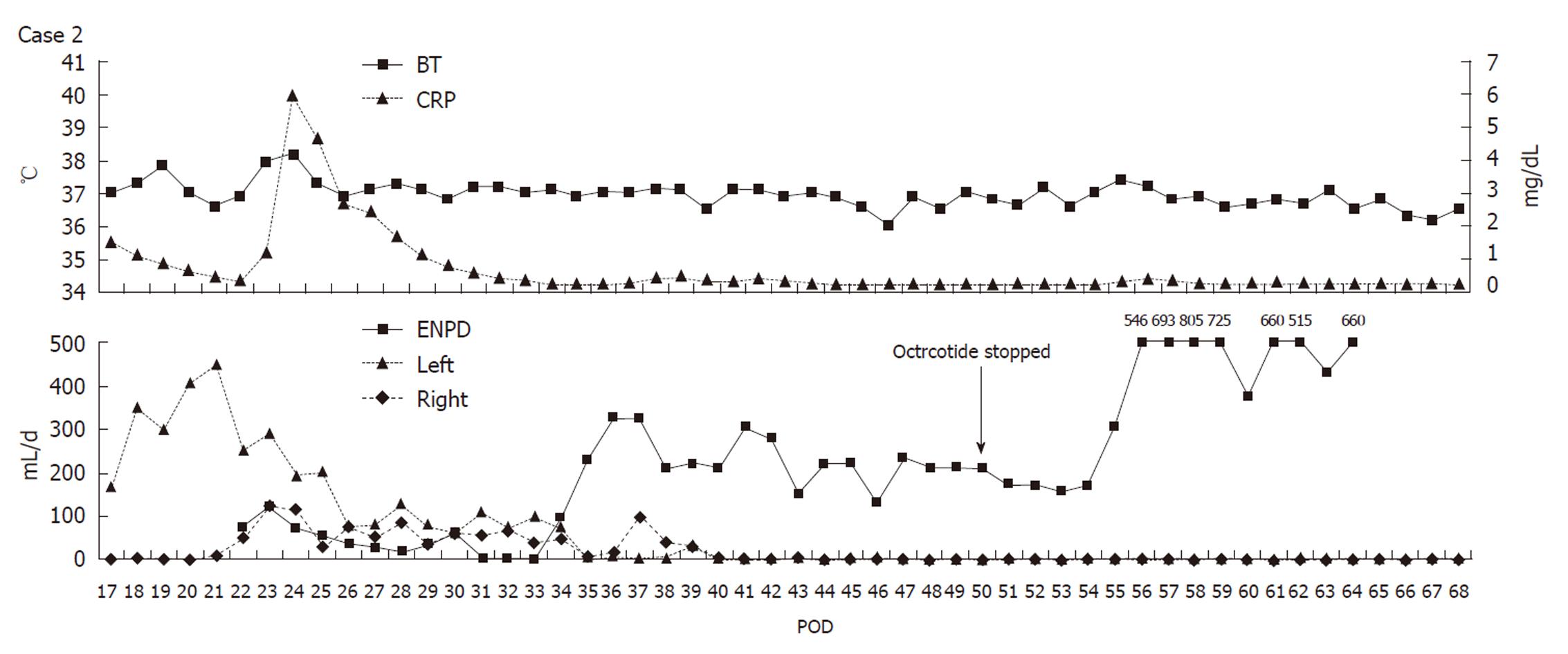
Figure 8 Upper chart shows the body temperature and serum C-reactive protein level.
Lower one shows daily output of endoscopic naso-pancreatic drainage (ENPD) tube and the drains at both the edges of the pancreas (Right and Left) in case 2. The pancreatic fistula healed completely on post-ENPD day 38. BT: Body temperature; CRP: C-reactive protein; POD: Postoperative day.
Figure 9 Computed tomography performed 14 d after removal of the drains in case 2 showing no fluid collection around the pancreas.
- Citation: Nagatsu A, Taniguchi M, Shimamura T, Suzuki T, Yamashita K, Kawakami H, Abo D, Kamiyama T, Furukawa H, Todo S. Endoscopic naso-pancreatic drainage for the treatment of pancreatic fistula occurring after LDLT. World J Gastroenterol 2011; 17(30): 3560-3564
- URL: https://www.wjgnet.com/1007-9327/full/v17/i30/3560.htm
- DOI: https://dx.doi.org/10.3748/wjg.v17.i30.3560


