Copyright
©2011 Baishideng Publishing Group Co.
World J Gastroenterol. Aug 14, 2011; 17(30): 3487-3496
Published online Aug 14, 2011. doi: 10.3748/wjg.v17.i30.3487
Published online Aug 14, 2011. doi: 10.3748/wjg.v17.i30.3487
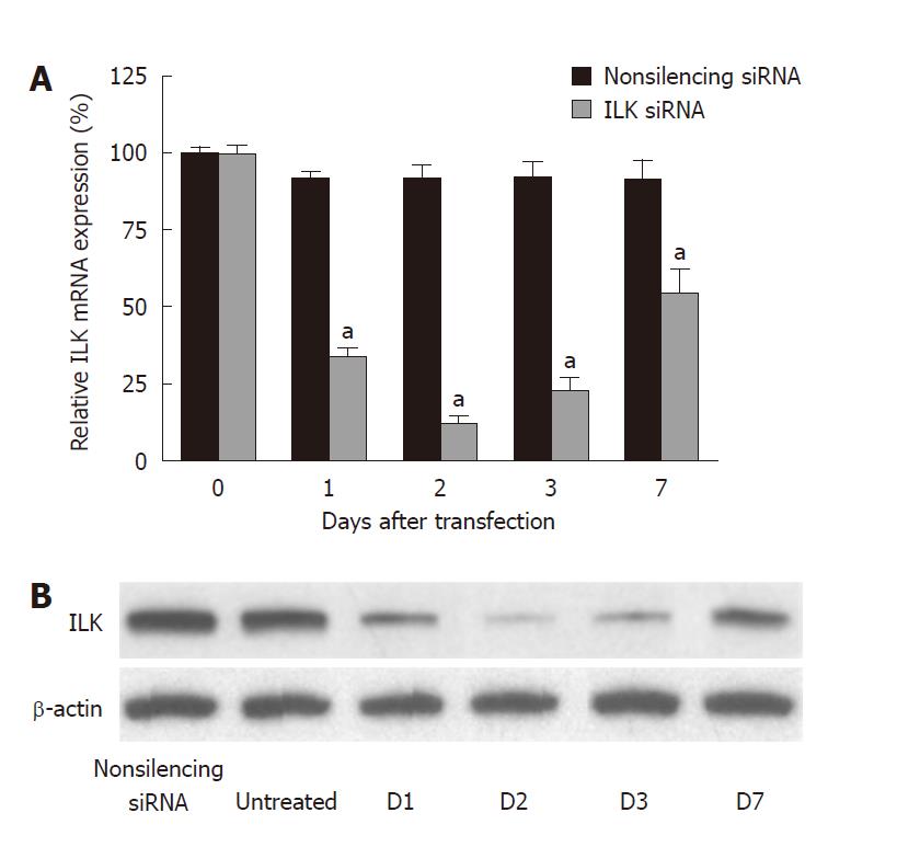
Figure 1 Effects of siRNA on integrin-linked kinase expression in gastric cancer cells.
Integrin-linked kinase expression levels were analyzed by (A) real-time polymerase chain reaction and (B) Western blot analysis at different time points after incubation. Data are the mean ± SD. aP < 0.05 vs control. Non-silencing siRNA is set to 100%.
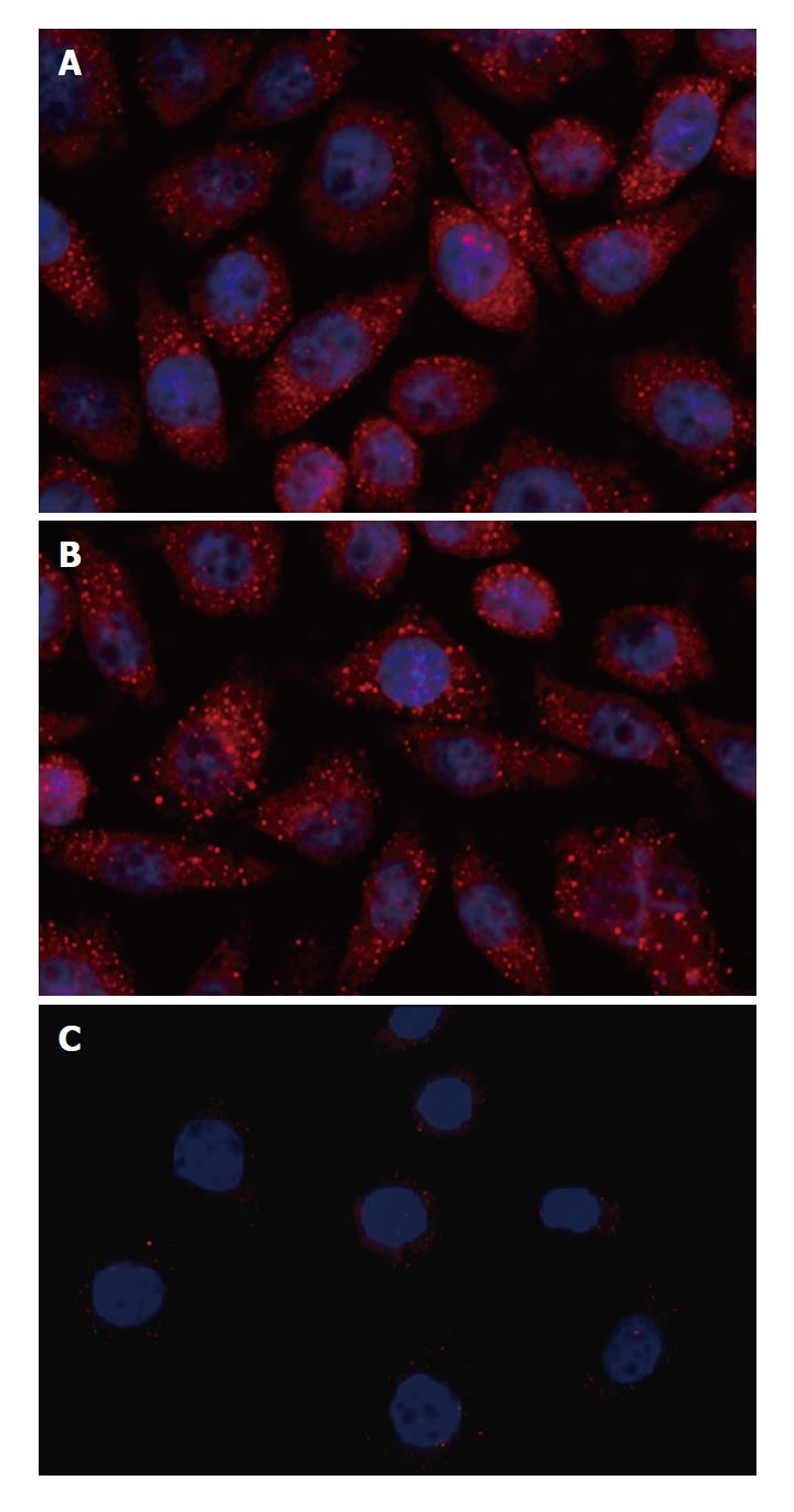
Figure 2 Immunocytochemical assays for integrin-linked kinase in gastric cancer cells.
The fluorescence intensity represents the expression of integrin-linked kinase (ILK) in gastric cancer cells. A: Untransfected cells (Untreated); B: Non-silencing siRNA-transfected cells (Non-silencing siRNA); C: ILK siRNA-transfected cells (ILK siRNA). Experiments were repeated three times.
Figure 3 Effects of integrin-linked kinase on attachment of gastric cancer cells.
Cell attachment was assessed after 1 h incubation and subsequent MTT test. Data are mean ± SD of three independent experiments (bP < 0.01 vs control). Non-silencing siRNA is set to 100%. ILK: Integrin-linked kinase.
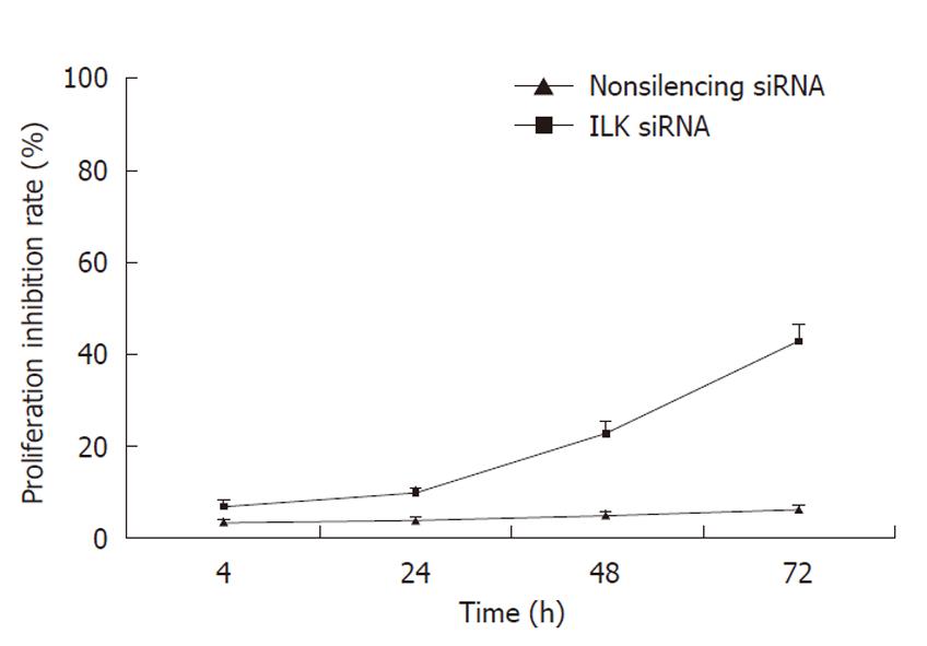
Figure 4 Effects of integrin-linked kinase on proliferation of gastric cancer cells.
Gastric cancer BGC-823 cells were treated with integrin-linked kinase siRNA or non-silencing siRNA. Six duplicate wells were set up for each sample. After 4, 24, 48 or 72 h incubation, 20 μL MTT (5 g/L) was added to each well. Data are the mean ± SD of three independent experiments. Cell proliferation is plotted against time.
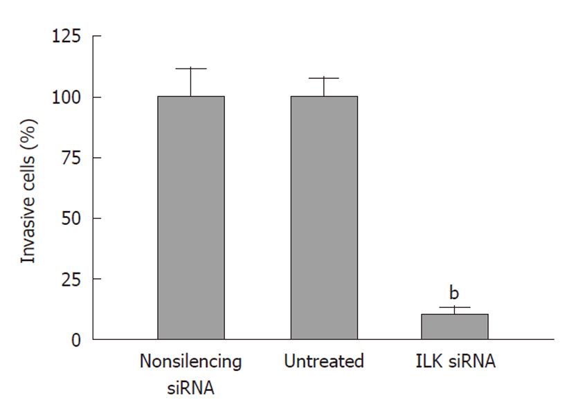
Figure 5 Effects of integrin-linked kinase on invasion of gastric cancer cells.
Cell invasion was assessed 24 h after incubation in Matrigel coated transwell culture cambers. Data are the mean ± SD of three independent experiments (bP < 0.01 vs control). Non-silencing siRNA is set to 100%. ILK: Integrin-linked kinase.
Figure 6 Effects of integrin-linked kinase on microfilament dynamics of gastric cancer cells.
Microfilament organization was assessed by immunofluorescence analysis with rhodamine-phalloidin. A, B: Non-silencing siRNA-transfected cells displayed an elaborate network of precisely organized F-actin filaments and a high degree of cell spreading; C, D: The F-actin filament architecture became significantly disturbed with less lamellipodia and filopodia formation in integrin-linked kinase knockdown cells compared with control cells. Experiments were repeated three times.
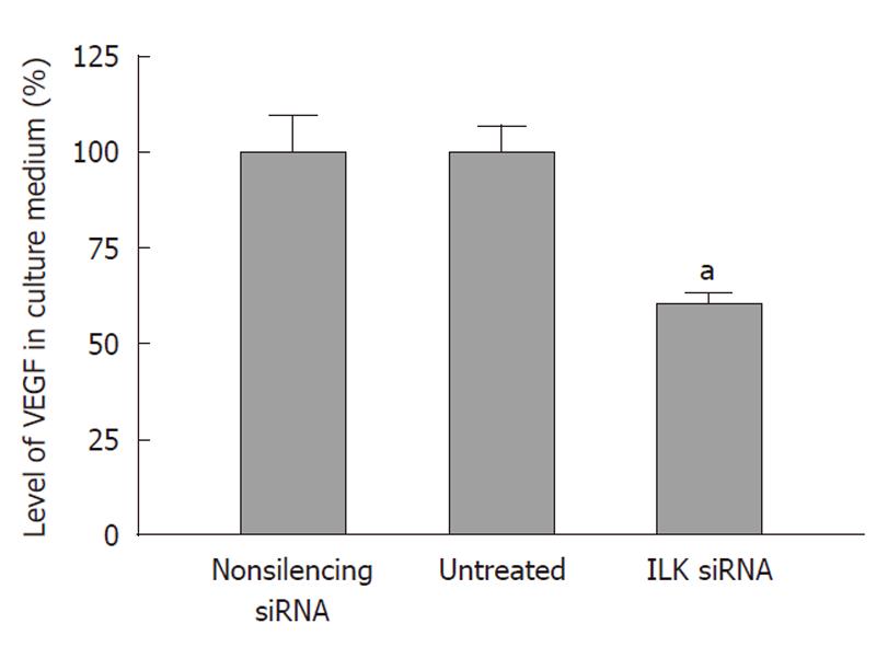
Figure 7 Effects of integrin-linked kinase on vascular endothelial growth factor secretion by gastric cancer cells.
After transfection, the culture medium was harvested and cell counting was performed. Vascular endothelial growth factor (VEGF) released into the culture supernatant was measured by enzyme-linked immunosorbent assay. Data are the mean ± SD of three independent experiments (aP < 0.05 vs control). Non-silencing siRNA cells are set to 100%. ILK: Integrin-linked kinase.
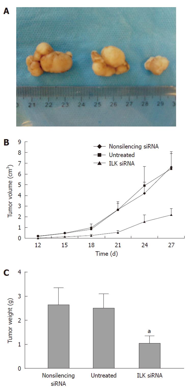
Figure 8 Integrin-linked kinase siRNA inhibits the growth of gastric cancer xenografts in nude mice.
BGC-823 cells were treated with integrin-linked kinase (ILK) siRNA or non-silencing siRNA or left untreated. Equal numbers of cells (1 × 10⁷) were injected 48 h later subcutaneously into the armpit of five nude mice in each group. Tumor formation was scored every 3 d. At week 4 after injection, the mice were killed and tumor weight recorded. A: images of tumors excised from the nude mice; B: estimated tumor volume of non-silencing siRNA, untreated or siRNA ILK tumors; C: estimated tumor weights of non-silencing siRNA, untreated or siRNA ILK tumors (aP < 0.05 vs control).
- Citation: Zhao G, Guo LL, Xu JY, Yang H, Huang MX, Xiao G. Integrin-linked kinase in gastric cancer cell attachment, invasion and tumor growth. World J Gastroenterol 2011; 17(30): 3487-3496
- URL: https://www.wjgnet.com/1007-9327/full/v17/i30/3487.htm
- DOI: https://dx.doi.org/10.3748/wjg.v17.i30.3487


