Copyright
©2010 Baishideng Publishing Group Co.
World J Gastroenterol. Dec 28, 2010; 16(48): 6139-6144
Published online Dec 28, 2010. doi: 10.3748/wjg.v16.i48.6139
Published online Dec 28, 2010. doi: 10.3748/wjg.v16.i48.6139
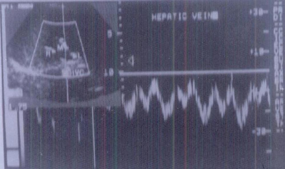
Figure 1 Triphasic waveform pattern of normal hepatic vein.
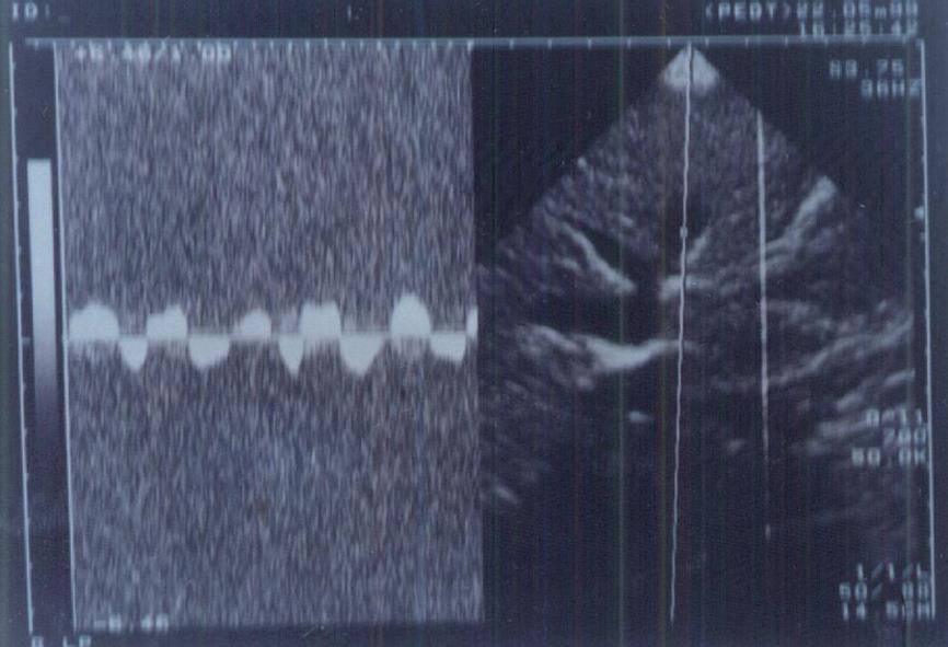
Figure 2 Abnormal waveform pattern of hepatic veins in a cirrhotic liver.
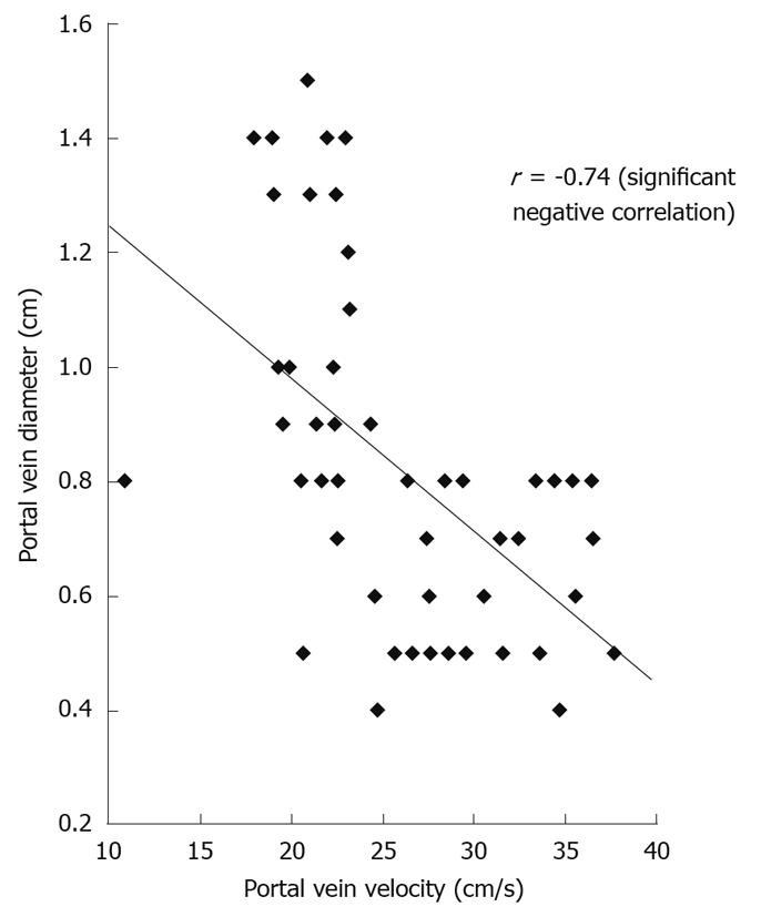
Figure 3 Correlation between portal vein flow velocity and portal vein diameter.
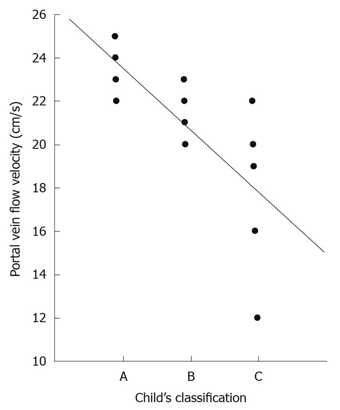
Figure 4 Correlation between Child-Pugh’s classification and portal vein velocity.
- Citation: El-Shabrawi MH, El-Raziky M, Sheiba M, El-Karaksy HM, El-Raziky M, Hassanin F, Ramadan A. Value of duplex doppler ultrasonography in non-invasive assessment of children with chronic liver disease. World J Gastroenterol 2010; 16(48): 6139-6144
- URL: https://www.wjgnet.com/1007-9327/full/v16/i48/6139.htm
- DOI: https://dx.doi.org/10.3748/wjg.v16.i48.6139


