Copyright
©2010 Baishideng Publishing Group Co.
World J Gastroenterol. Dec 14, 2010; 16(46): 5901-5906
Published online Dec 14, 2010. doi: 10.3748/wjg.v16.i46.5901
Published online Dec 14, 2010. doi: 10.3748/wjg.v16.i46.5901
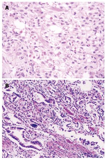
Figure 1 Hematoxylin and eosin staining in the concurrent esophageal and gastric cardia cancers tissue specimens.
A: Esophageal squamous cell carcinoma tissue (× 100); B: Gastric cardia adenocarcinoma tissue (× 100).
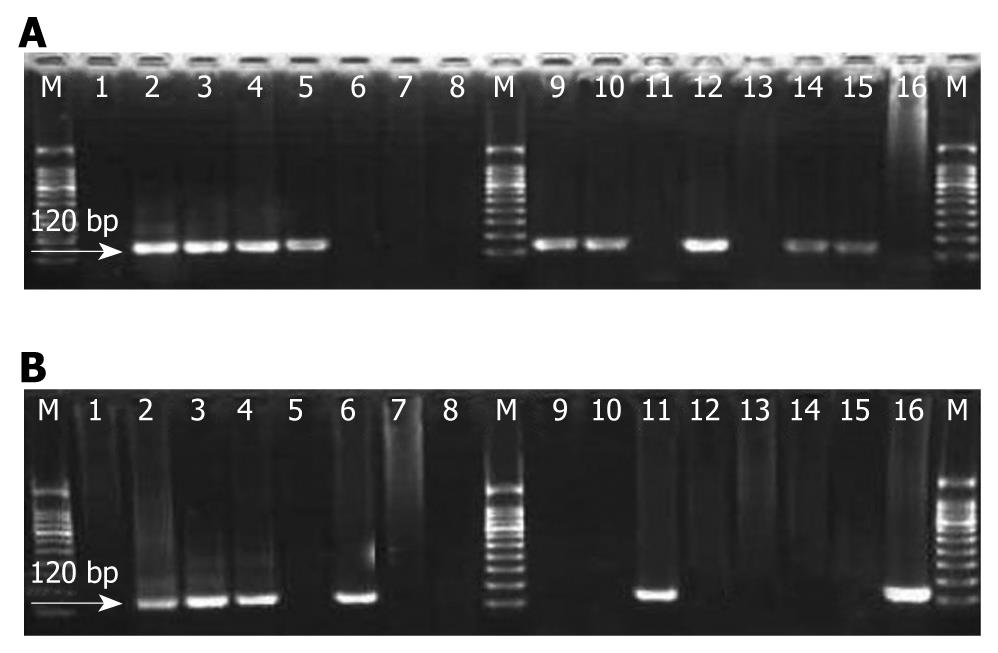
Figure 2 Amplification of human papillomavirus type 16-E6 gene fragment in the concurrent esophageal and gastric cardia cancers tissues.
Polymerase chain reaction products were run in 3.0% agarose gel; Lane M: Molecular marker (100 bp ladder); Lane 1: Double water (negative control); Lane 2: Plasmid with human papillomavirus type 16 (HPV16)-E6 120 bp (positive control); Lanes 3-16: Represent positive and negative cases. A: HPV16-E6 gene fragment amplification in esophageal squamous cell carcinoma tissues; B: HPV16-E6 gene fragment in the corresponding gastric cardia adenocarcinoma tissue amplification.
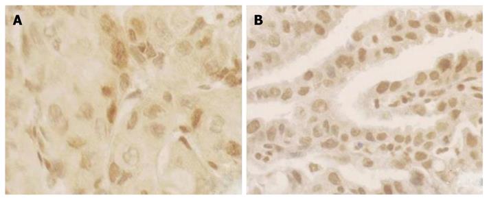
Figure 3 P16INK4A protein expression in the concurrent esophageal and gastric cardia cancers tissues by immunohistochemical staining Avidin-Biotin-Peroxidase Complex method (× 200).
A: P16INK4A protein expression in esophageal squamous cell carcinoma; B: Expression of P16INK4A protein in gastric cardia adenocarcinoma.
- Citation: Ding GC, Ren JL, Chang FB, Li JL, Yuan L, Song X, Zhou SL, Guo T, Fan ZM, Zeng Y, Wang LD. Human papillomavirus DNA and P16INK4A expression in concurrent esophageal and gastric cardia cancers. World J Gastroenterol 2010; 16(46): 5901-5906
- URL: https://www.wjgnet.com/1007-9327/full/v16/i46/5901.htm
- DOI: https://dx.doi.org/10.3748/wjg.v16.i46.5901


