Copyright
©2010 Baishideng Publishing Group Co.
World J Gastroenterol. Aug 28, 2010; 16(32): 4100-4106
Published online Aug 28, 2010. doi: 10.3748/wjg.v16.i32.4100
Published online Aug 28, 2010. doi: 10.3748/wjg.v16.i32.4100
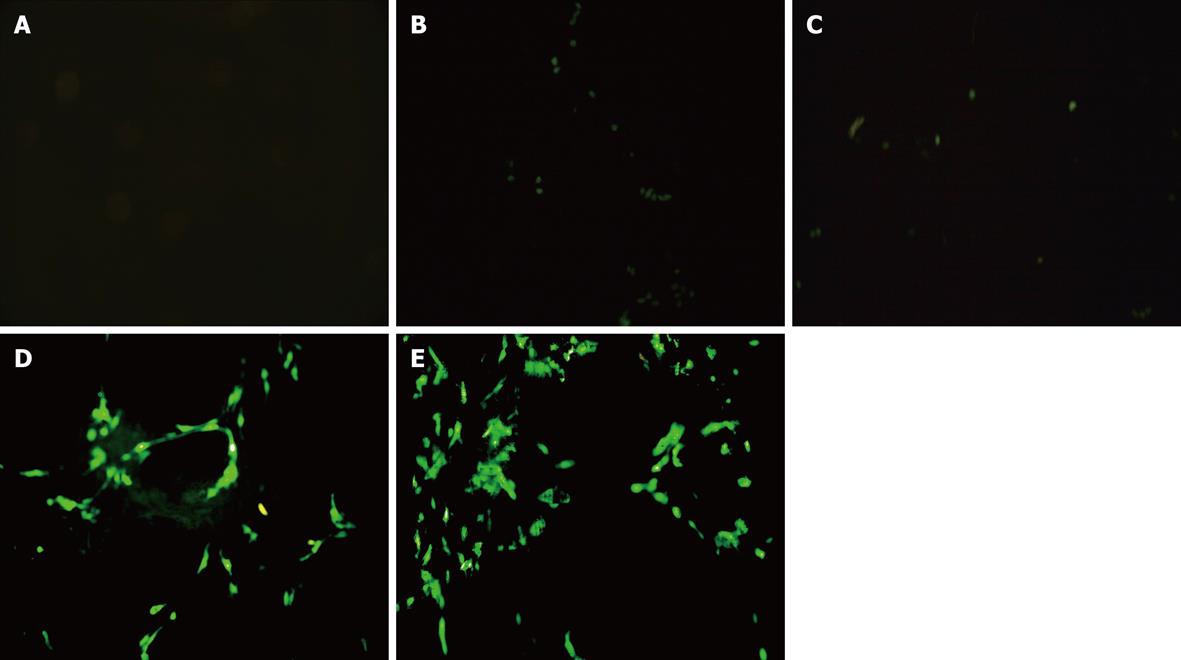
Figure 1 Expression of enhanced green fluorescent protein at 48 h after treatment (fluorescent images, original magnification × 200).
A: Control group; B: Fibronectin group; C: Sofast group; D: HK group; E: Focal adhesion kinase (FAK) short hairpin RNA group. FAK short hairpin RNA plasmids were successfully transfected into hepatic stellate cells. The results from fluorescence microscopy and flow cytometry showed that the transfection efficiency was 40% at 48 h.
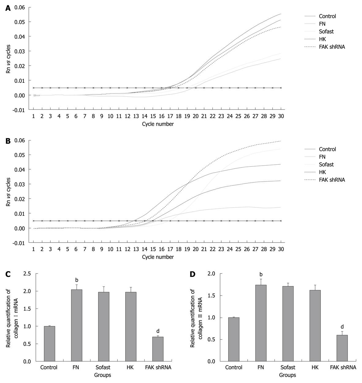
Figure 2 Focal adhesion kinase short hairpin RNA selectively inhibited the expressions of collagen I mRNA and collagen III mRNA in hepatic stellate cells after focal adhesion kinase short hairpin RNA transfection.
A, B: Real-time polymerase chain reaction SYBR Green I fluorescence history vs cycle number of target gene 1 (collagen I, A) and target gene 2 (collagen III, B) in sample cDNA. The cycle threshold (Ct) is shown by the darker horizontal line; C, D: The relative quantification of collagen I mRNA (C) and collagen III mRNA (D) are calculated according to 2-ΔΔC(t), [ΔΔCt = (CtCollagen I or III - CtGAPDH) experimental group - (CtCollagen I or III - CtGAPDH) control group] and shown in the bar graphs (n = 3, bP < 0.01 vs control, dP < 0.01 vs HK). It showed that the levels of type I collagen and type III collagen mRNA transcripts in fibronectin (FN) group was significantly higher than in the control group. FAK: Focal adhesion kinase; shRNA: Short hairpin RNA.
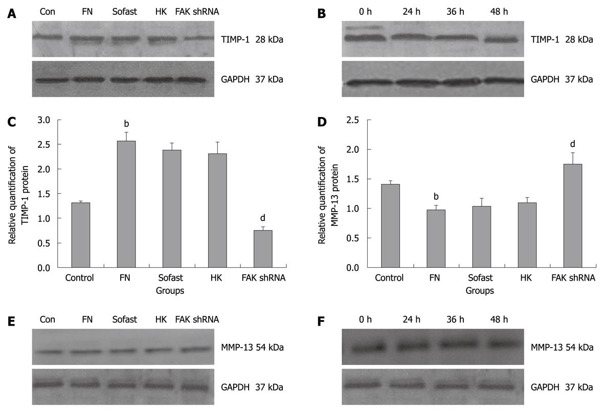
Figure 3 Focal adhesion kinase short hairpin RNA specifically inhibits the expressions of tissue inhibitors of metalloproteinases-1 protein and promotes the expressions of matrix metalloproteinases-13 protein in hepatic stellate cells.
A: Cells were harvested, lysed and total protein extracts were separated by SDS-PAGE and analyzed by Western blotting with polyclonal anti-tissue inhibitors of metalloproteinases-1 (TIMP-1) antibody. GAPDH served as a loading control; B: Western blotting analysis was used to detect the expressions of TIMP-1 at different time points; C: TIMP-1 expression levels obtained from scanning densitometry were expressed as a ratio of integral optical density value (IOD) TIMP-1/IOD GAPDH (n = 3, bP < 0.01 vs Con, dP < 0.01 vs HK); D: Matrix metalloproteinases-13 (MMP-13) expression levels obtained from scanning densitometry were expressed as a ratio of IOD MMP-13/IOD GAPDH (n = 3, bP < 0.01 vs Con, dP < 0.01 vs HK); E, F: Western blotting analysis was carried out at different groups (E) and different time points (F) using polyclonal anti-MMP-13 antibody and monoclonal anti-GAPDH antibody. FN: Fibronectin; FAK: Focal adhesion kinase; shRNA: Short hairpin RNA.
-
Citation: Dun ZN, Zhang XL, An JY, Zheng LB, Barrett R, Xie SR. Specific shRNA targeting of
FAK influenced collagen metabolism in rat hepatic stellate cells. World J Gastroenterol 2010; 16(32): 4100-4106 - URL: https://www.wjgnet.com/1007-9327/full/v16/i32/4100.htm
- DOI: https://dx.doi.org/10.3748/wjg.v16.i32.4100


