Copyright
©2009 The WJG Press and Baishideng.
World J Gastroenterol. Feb 14, 2009; 15(6): 761-763
Published online Feb 14, 2009. doi: 10.3748/wjg.15.761
Published online Feb 14, 2009. doi: 10.3748/wjg.15.761
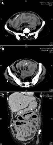
Figure 1 CT showing pronounced contrast enhancement of peritoneum and thickened intestinal wall.
A: Most nodules coalesced to form large omental plaques (omental cakes); B: A moderate amount of ascites located between intestinal canals; C: No actual large abdominal masses.
- Citation: Hou XQ, Cui HH, Jin X. Coexistence of tuberculous peritonitis and primary papillary serous carcinoma of the peritoneum: A case report and review of the literature. World J Gastroenterol 2009; 15(6): 761-763
- URL: https://www.wjgnet.com/1007-9327/full/v15/i6/761.htm
- DOI: https://dx.doi.org/10.3748/wjg.15.761


