Copyright
©2009 The WJG Press and Baishideng.
World J Gastroenterol. Oct 28, 2009; 15(40): 5106-5108
Published online Oct 28, 2009. doi: 10.3748/wjg.15.5106
Published online Oct 28, 2009. doi: 10.3748/wjg.15.5106
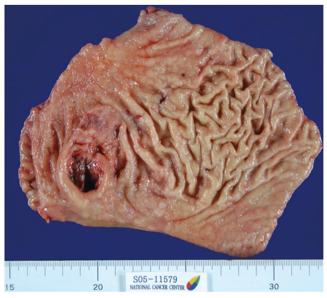
Figure 1 Gross pathology showed a 5.
8 cm × 3.2 cm ulcerofungating mass in the antrum, with extensive hemorrhage and light gray fibrosis, and a 2.5 cm × 2.0 cm ulcerative lesion.
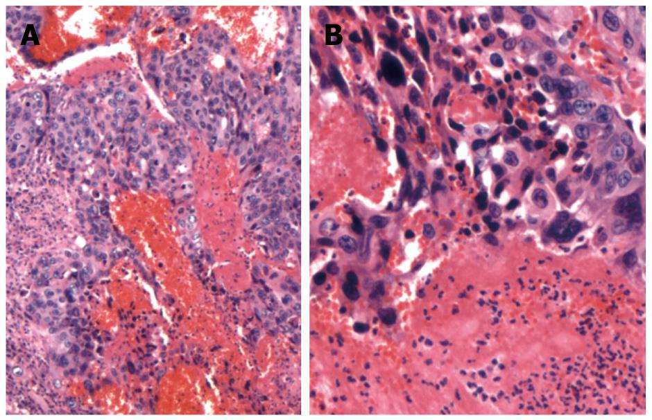
Figure 2 Microscopically, massive numbers of pleomorphic, bizarre tumor cells with hemorrhage were revealed: syncytiotrophoblasts and cytotrophoblasts (HE, A × 40, B × 100).
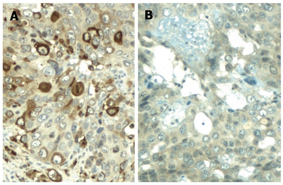
Figure 3 Immunohistochemical staining showed positive immunoreactivity for β-human chorionic gonadotropin (A) and focal positivity for α-fetoprotein (B).
- Citation: Eom BW, Jung SY, Yoon H, Kook MC, Ryu KW, Lee JH, Kim YW. Gastric choriocarcinoma admixed with an α-fetoprotein-producing adenocarcinoma and separated adenocarcinoma. World J Gastroenterol 2009; 15(40): 5106-5108
- URL: https://www.wjgnet.com/1007-9327/full/v15/i40/5106.htm
- DOI: https://dx.doi.org/10.3748/wjg.15.5106


