Copyright
©2009 The WJG Press and Baishideng.
World J Gastroenterol. Oct 21, 2009; 15(39): 4952-4957
Published online Oct 21, 2009. doi: 10.3748/wjg.15.4952
Published online Oct 21, 2009. doi: 10.3748/wjg.15.4952
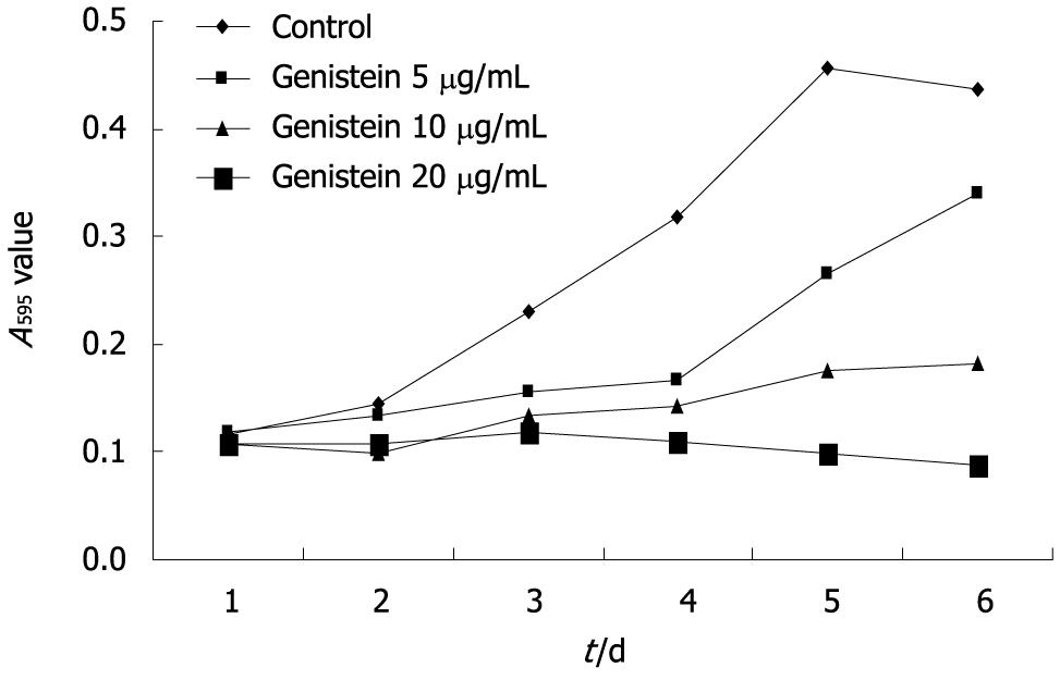
Figure 1 Inhibitory effects of genistein on MHCC97-H cell proliferation.
MHCC97-H cells were treated with various concentrations of genistein for 6 d, control cells were left untreated, A595 values were determined daily.
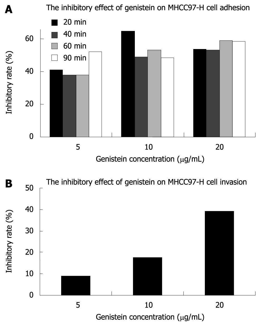
Figure 2 Inhibitory effects of genistein on adhesion and invasion of MHCC97-H cells.
A: MHCC97-H cells were incubated for 20, 40, 60 and 90 min with various concentrations of genistein; B: MHCC97-H cells were incubated for 20 h with various concentrations of genistein.
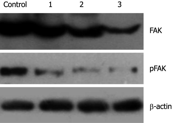
Figure 3 Western blotting analysis of protein expression and phosphorylation of FAK in MHCC97-H cells following genistein treatment.
Lane 1: Genistein 5 μg/mL; Lane 2: Genistein 10 μg/mL; Lane 3: Genistein 20 μg/mL.
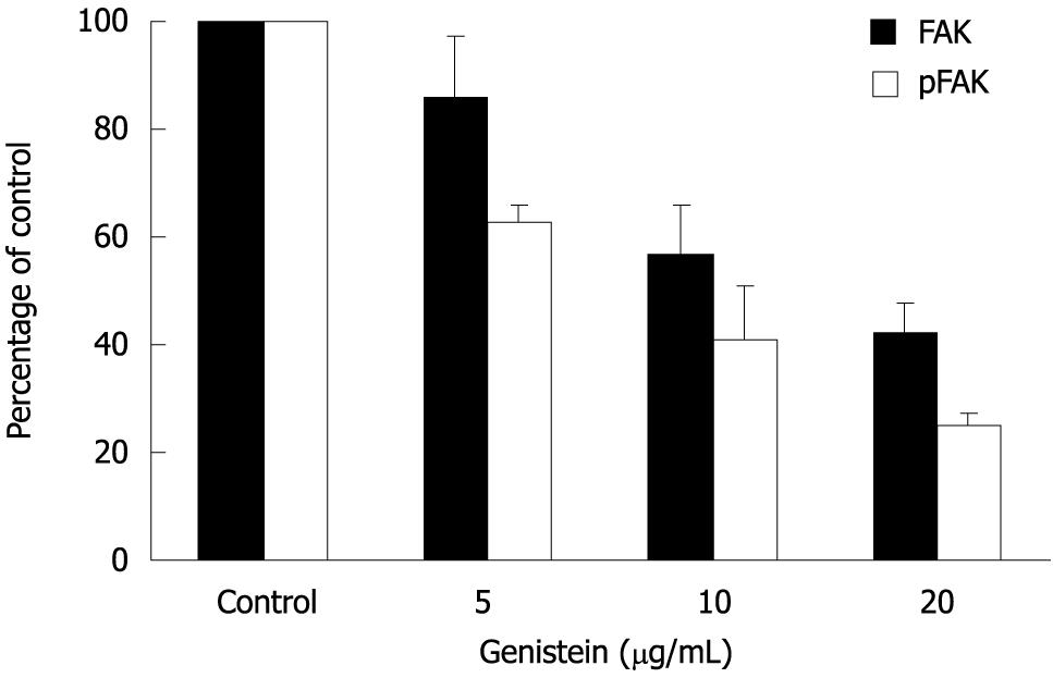
Figure 4 Effects of genistein on protein expression and phosphorylation of FAK.
Black columns, protein expression of FAK; Open columns, phosphorylation of FAK. Values are mean ± SD from experiments.
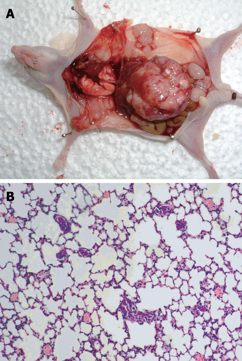
Figure 5 Autopsy and microphotographic appearance after orthotopic implantation.
A: Autopsy of mice bearing orthotopic implantations. Rapid tumor growth occurred. Thirty-five days after tumor tissue implantation into the liver; B: Microphotography of lung metastasis by MHCC97-H cells. Thirty-five days after orthotopic implantation, MHCC97-H cells metastasized to the lungs, forming pulmonary metastatic lesions. HE, × 200.
- Citation: Gu Y, Zhu CF, Dai YL, Zhong Q, Sun B. Inhibitory effects of genistein on metastasis of human hepatocellular carcinoma. World J Gastroenterol 2009; 15(39): 4952-4957
- URL: https://www.wjgnet.com/1007-9327/full/v15/i39/4952.htm
- DOI: https://dx.doi.org/10.3748/wjg.15.4952


