Copyright
©2009 The WJG Press and Baishideng.
World J Gastroenterol. Apr 21, 2009; 15(15): 1880-1885
Published online Apr 21, 2009. doi: 10.3748/wjg.15.1880
Published online Apr 21, 2009. doi: 10.3748/wjg.15.1880
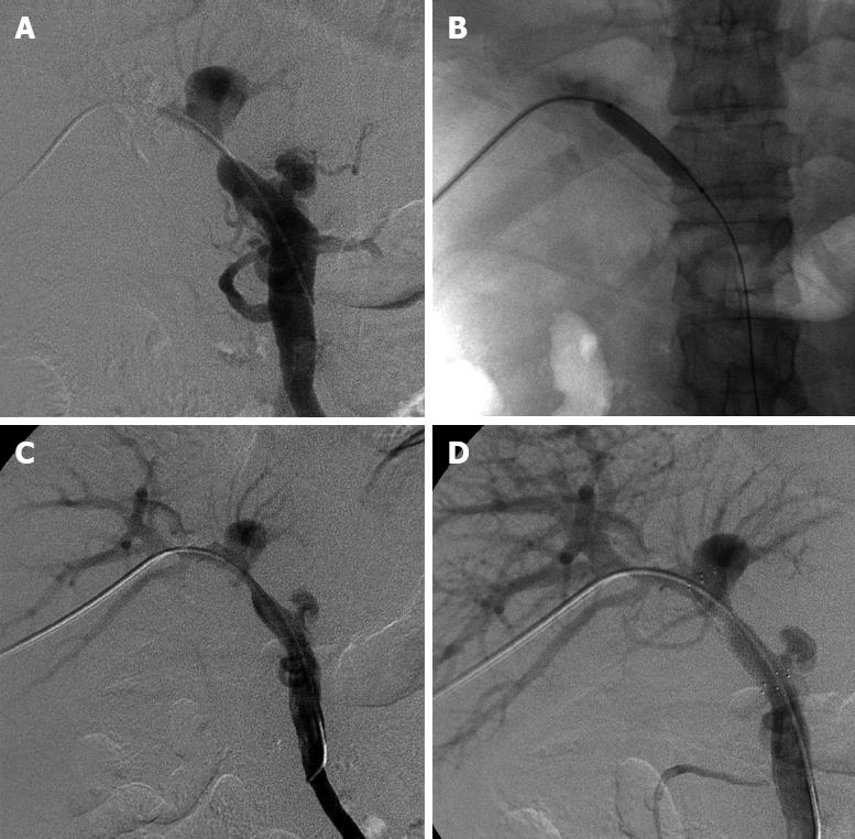
Figure 1 Procedure of percutaneous transhepatic portal venoplasty and stenting.
A: Percutaneous transhepatic portography revealed a severe anastomotic stenosis and gastroesophageal varices; B: Portal venoplasty was performed with an undersized balloon and the balloon was slowly inflated; C: Postvenoplasty portography showed a residual stenosis, but gastroesophageal varices became almost invisible; D: Poststenting portography visualized portal venous patency and inflow had recovered completely.
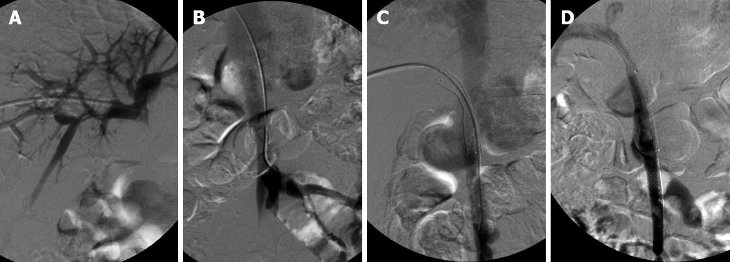
Figure 2 A 28-year-old man with a pre-existing meso-caval shunt.
A: Initial portography revealed an almost occlusive anastomosis; B: Portography detected a pre-existing meso-caval shunt due to visualization of the inferior vena cava; C: After stenting, the contrast agents flowed mainly into the inferior vena cava via the shunt and little flowed into the portal vein; D: A covered stent was deployed into the superior mesenteric vein to occlude the shunt. Portal hepatopetal flow was restored and the inferior vena cava became invisible.
- Citation: Wei BJ, Zhai RY, Wang JF, Dai DK, Yu P. Percutaneous portal venoplasty and stenting for anastomotic stenosis after liver transplantation. World J Gastroenterol 2009; 15(15): 1880-1885
- URL: https://www.wjgnet.com/1007-9327/full/v15/i15/1880.htm
- DOI: https://dx.doi.org/10.3748/wjg.15.1880


