Copyright
©2008 The WJG Press and Baishideng.
World J Gastroenterol. Feb 7, 2008; 14(5): 776-781
Published online Feb 7, 2008. doi: 10.3748/wjg.14.776
Published online Feb 7, 2008. doi: 10.3748/wjg.14.776
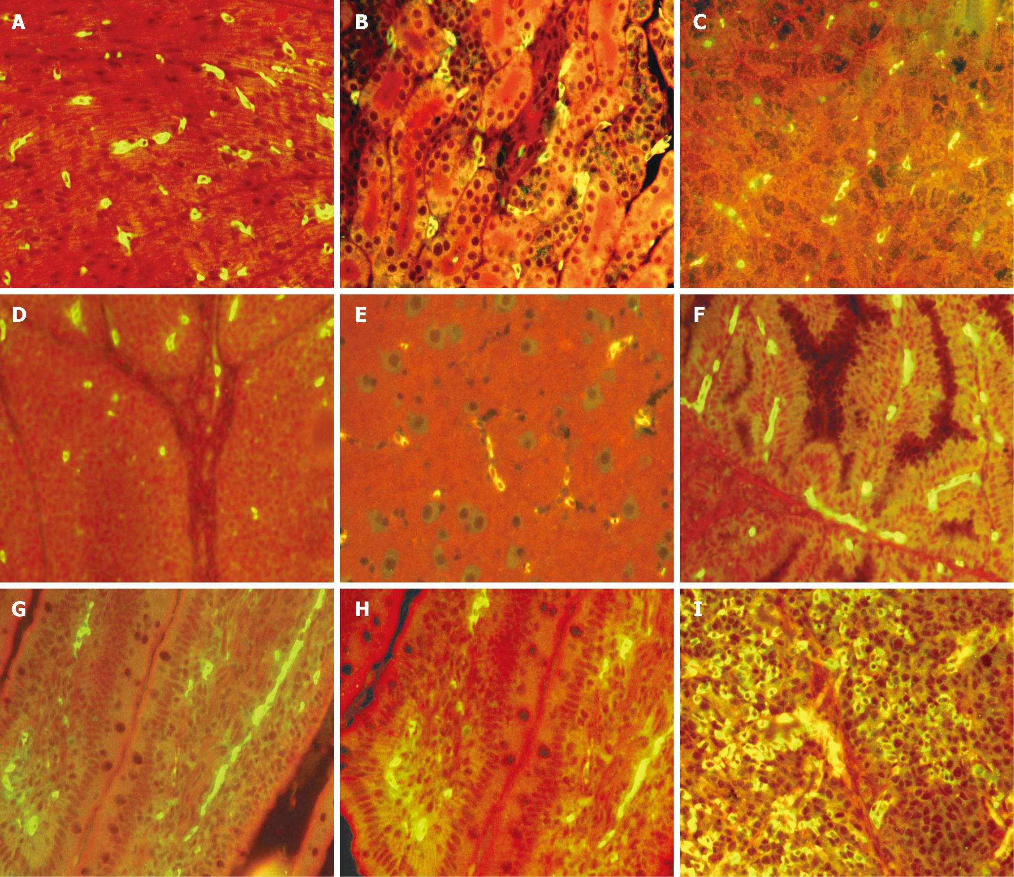
Figure 1 Organ tissues from SE-inoculated ducks immunofluorescent stain for SE.
A: Positive staining baclli are adhering to the Cardiac muscle fiber (Heart); B: Positive staining baclli are adhering to the mesenchyme between the tube of Kidney (Kidney); C: Positive staining baclli are adhering to the mesenchyme between the Hepatic cord; D: Positive staining baclli are adhering to the area of medulla of the Follicle; E: Positive staining baclli are adhering to vascular endothelial cell; F: Positive staining baclli are adhering to the mesenchyme of the tube of the gland; G, H: Positive staining bacllis are adhering to the interstitial tissue of the lamina propria in villi; I: Positive staining baclli are adhering to red medulla. (Images were acquired by using 40 x objective).
-
Citation: Yan B, Cheng AC, Wang MS, Deng SX, Zhang ZH, Yin NC, Cao P, Cao SY. Application of an indirect immunofluorescent staining method for detection of
Salmonella enteritidis in paraffin slices and antigen location in infected duck tissues. World J Gastroenterol 2008; 14(5): 776-781 - URL: https://www.wjgnet.com/1007-9327/full/v14/i5/776.htm
- DOI: https://dx.doi.org/10.3748/wjg.14.776


