Copyright
©2008 The WJG Press and Baishideng.
World J Gastroenterol. Dec 14, 2008; 14(46): 7075-7085
Published online Dec 14, 2008. doi: 10.3748/wjg.14.7075
Published online Dec 14, 2008. doi: 10.3748/wjg.14.7075
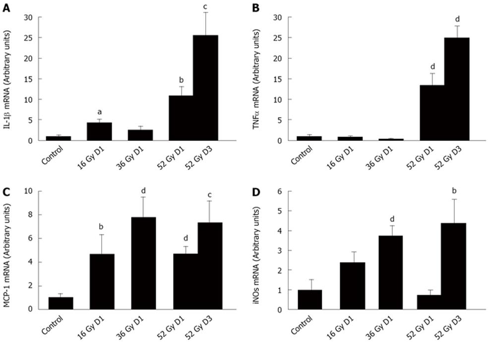
Figure 1 Effects of colorectal fractionated irradiation protocol on inflammatory mediator expression.
Levels of IL-1β (A), TNFα (B), MCP-1 (C) and iNOS (D) mRNA were assessed in distal colon mucosa one day after cumulative doses of 16 Gy, 36 Gy, 52 Gy as well as 3 d after 52 Gy. The quantification of target genes was normalized to the reference gene HPRT. Data are mean ± SE, aP < 0.05; bP < 0.01; cP < 0.005; dP < 0.001 vs control.
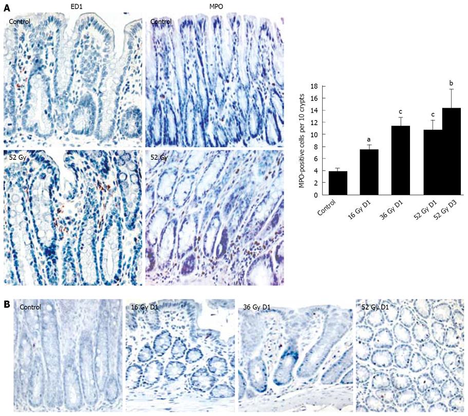
Figure 2 Effects of colorectal fractionated irradiation protocol on inflammatory cells and apoptotic cell presence.
Recruitment of ED-1-positive and MPO-positive cells (A) was assessed in distal colon mucosa in the normal distal colon (control) and three days after cumulative doses of 52 Gy. MPO-positive cells were correlated with the average number of neutrophils one day after cumulative doses of 16 Gy, 36 Gy, and one day and three days after 52 Gy. The presence of apoptotic cells was confirmed by the terminal deoxynucleotidyltransferase (TdT)-mediated dUTP-biotin nick-end labeling (TUNEL) staining at each timepoint of the fractionated protocol (B). Data are mean ± SE, aP < 0.01; bP < 0.005; cP < 0.001 vs control values. (× 20).
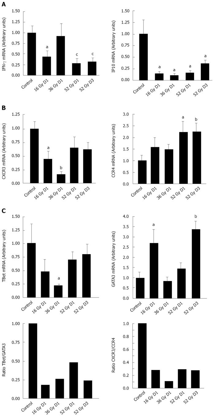
Figure 3 Early Th1/Th2 polarization by fractionated irradiation protocol within 1 and 3 d of irradiation.
Colorectal fractionated irradiation protocol induced suppression of Th1 cytokines (IFNγ and IFN-inducible genes (IP-10)) (A). Gene expression of the chemokine receptors CXCR3 and CCR4 (B) and transcription factors T-bet and Gata3 (C) implicated in the Th1/Th2 balance was analyzed one day after cumulative doses of 16 Gy, 36 Gy, 52 Gy and 3 d after 52 Gy in colon mucosa. Data are mean ± SE, aP < 0.05; bP < 0.01; cP < 0.005 vs control.
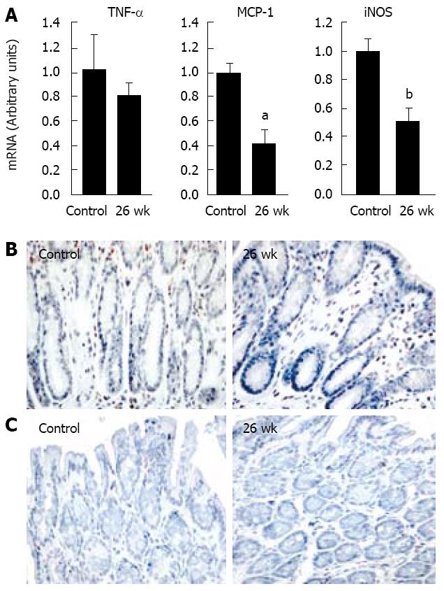
Figure 4 Long-term impairment of inflammatory response.
Delayed effects of the radiation schedule on expression of TNFα, MCP-1 and iNOS in colon mucosa 26 wk after the end of cumulative dose of 52 Gy (A). The quantification of target genes was normalized to the reference gene HPRT. Immunostaining for macrophages (B) and apoptotic cells (C) in the colon: Fractionated irradiated group shows no intense ED1-positive macrophages in the lamina propria and no significant modification of the apoptotic cell number 26 wk post-irradiation. Data are mean ± SE, aP < 0.01; bP < 0.005 vs control.
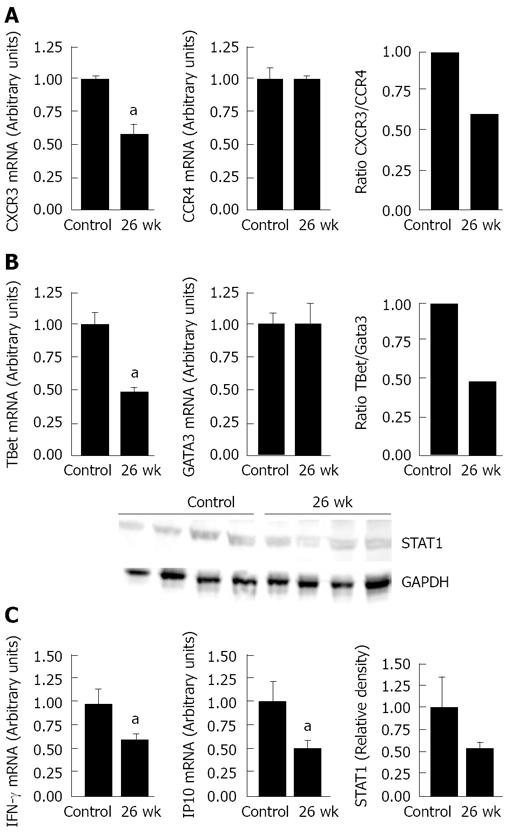
Figure 5 Persistence of Th2 polarization at 6 mo.
Expression of chemokine receptors CXCR3 and CCR4 (A) and transcription factors T-bet and Gata3 expression (B) in colon mucosa was assessed 26 wk after the end of cumulative dose of 52Gy. Aspects of immune response that changed long after irradiation included IFNγ and IP-10 expression and downregulation of signal transducer and activator 1 (STAT1) (C). Immunoblot showing STAT1 in total protein extracts from mucosa colon. GAPDH levels are used as internal standards and relative densitometric data are analysed following normalization to the control. Dot plots of four experiments on different samples are shown. Data are the mean ± SE; aP < 0.05 vs control.
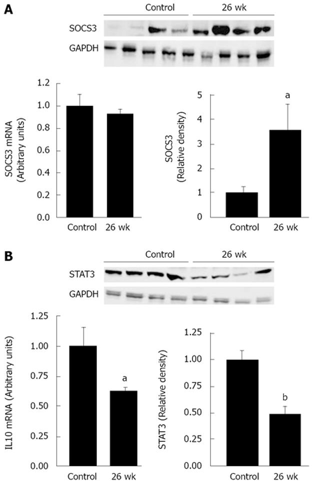
Figure 6 Fractionated irradiation-modified SOCS3, IL-10 and STAT3 expression.
SOCS3 mRNA and protein (A), IL-10 mRNA and STAT3 protein (B) were measured in the colonic mucosa 26 wk after the end of cumulative dose of 52 Gy. Western blot analyses are showing SOCS3 and STAT3 in total protein extracts from mucosa colon. GAPDH levels are used as internal standards and relative densitometric data are analysed following normalization to the control. Dot plots of four experiments on different samples are shown. Data are expressed as the mean ± SE; aP < 0.05; bP < 0.001 vs control.
- Citation: Grémy O, Benderitter M, Linard C. Acute and persisting Th2-like immune response after fractionated colorectal γ-irradiation. World J Gastroenterol 2008; 14(46): 7075-7085
- URL: https://www.wjgnet.com/1007-9327/full/v14/i46/7075.htm
- DOI: https://dx.doi.org/10.3748/wjg.14.7075


