Copyright
©2008 The WJG Press and Baishideng.
World J Gastroenterol. Aug 28, 2008; 14(32): 5008-5014
Published online Aug 28, 2008. doi: 10.3748/wjg.14.5008
Published online Aug 28, 2008. doi: 10.3748/wjg.14.5008
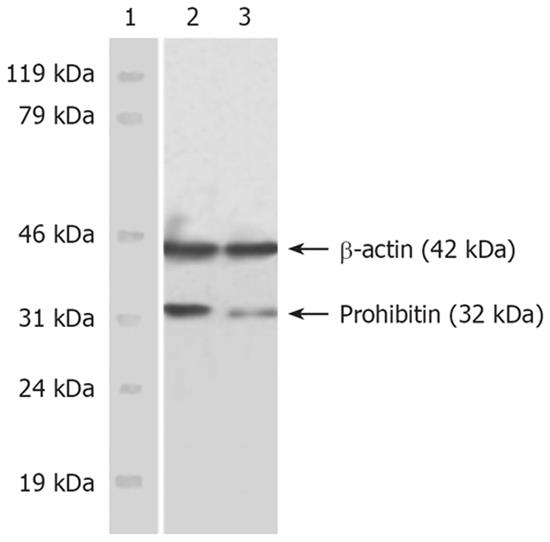
Figure 1 Western blot of PHB in nuclear matrix of SMMC-7721 cell.
Lane 1: Marker; Lane 2: SMMC-7721 cells; Lane 3: HMBA-treated cells.
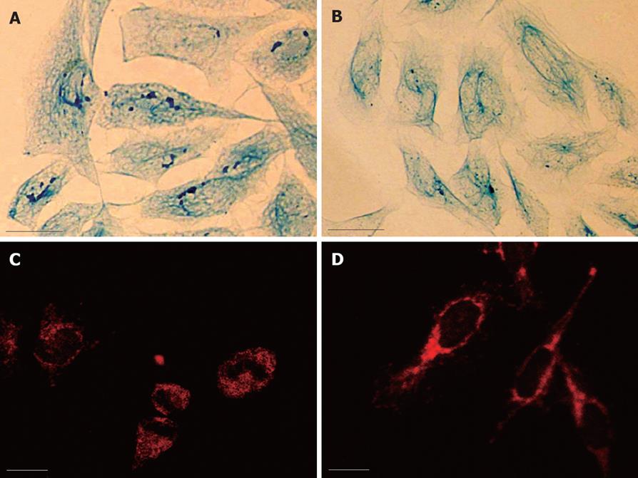
Figure 2 A: LM observation of NM-IF system in SMMC-7721 cells, coomassie brilliant blue staining, bar = 10 μm; B: LM observation of NM-IF system in SMMC-7721 cells after HMBA treatment, Coomassie brilliant blue staining, bar = 10 μm; C: Immunofluorescence staining of PHB in the NMIF system of SMMC-7721 cells, bar = 10 μm; D: Immunofluorescence staining of PHB in the nuclear matrix-intermediate filament system of SMMC-7721 cells after HMBA treatment, bar = 10 μm.
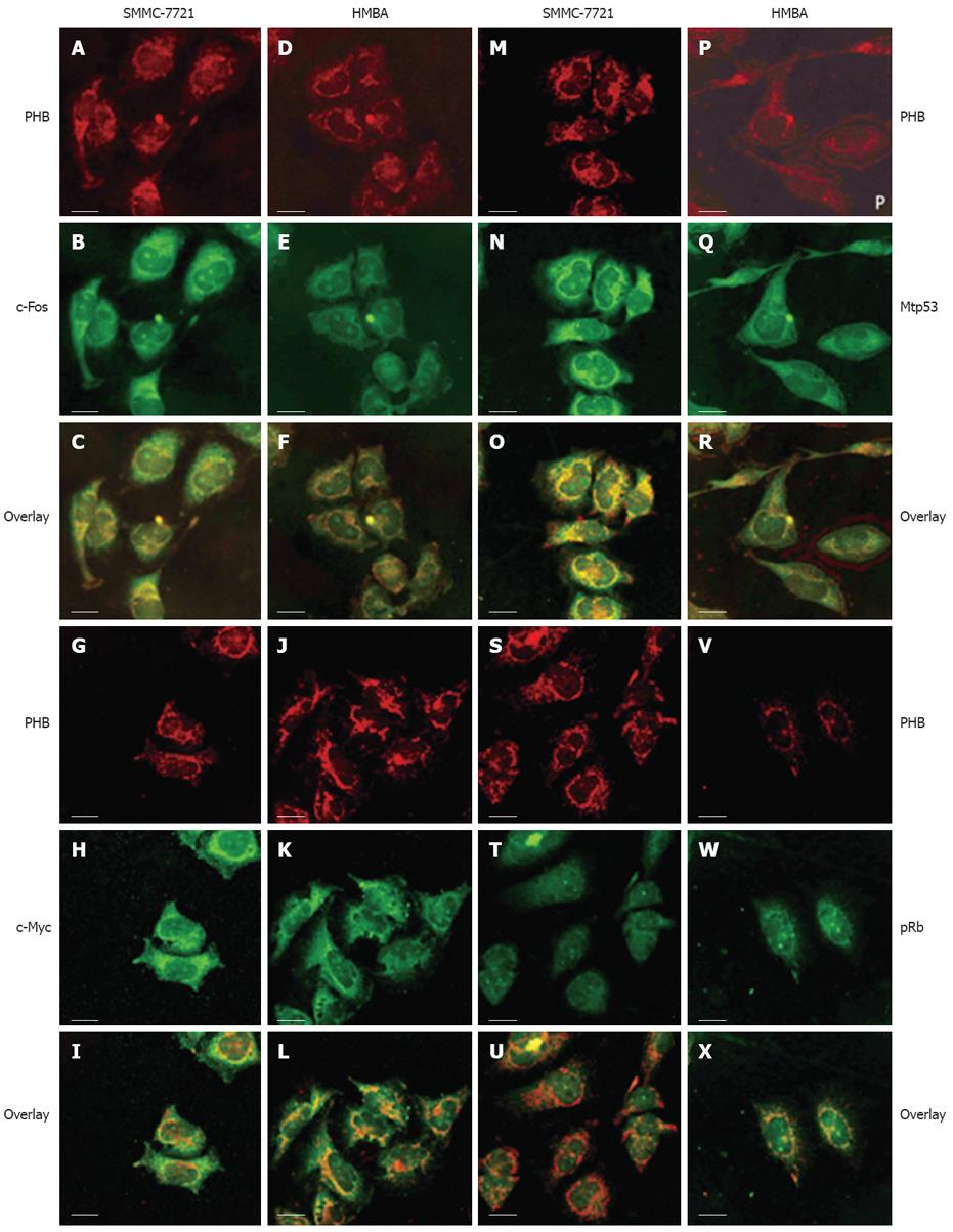
Figure 3 LSCM observation of the co-localization of PHB, bar = 10 μm.
A-F: With c-Fos in SMMC-7721 cells; G-L: With c-Myc in SMMC-7721 cells; M-R: With mtp53 in SMMC-7721 cells; S-X: With pRb in SMMC-7721 cells.
- Citation: Xu DH, Tang J, Li QF, Shi SL, Chen XF, Liang Y. Positional and expressive alteration of prohibitin during the induced differentiation of human hepatocarcinoma SMMC-7721 cells. World J Gastroenterol 2008; 14(32): 5008-5014
- URL: https://www.wjgnet.com/1007-9327/full/v14/i32/5008.htm
- DOI: https://dx.doi.org/10.3748/wjg.14.5008


