Copyright
©2008 The WJG Press and Baishideng.
World J Gastroenterol. Apr 14, 2008; 14(14): 2213-2217
Published online Apr 14, 2008. doi: 10.3748/wjg.14.2213
Published online Apr 14, 2008. doi: 10.3748/wjg.14.2213
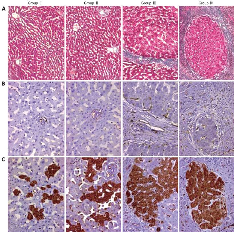
Figure 1 Liver fibrosis (A), angiogenesis (B) and TSP-1 expression (C) in the study group.
Liver fibrosis was stained by Masson trichrome at different time points of treatment and angiogenesis was evaluated with an anti-CD34 antibody. In normal livers, the number CD34 labeled vessels and TSP-1 positive cells is lower when compared to DEN treated livers. In the latter, their number increases according to the extent of fibrosis.
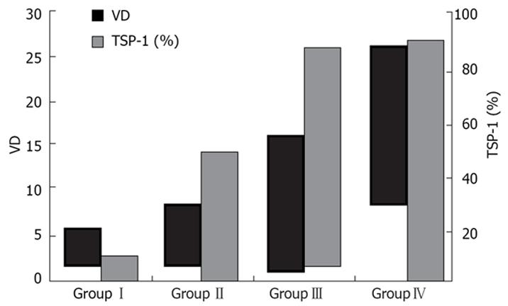
Figure 2 Results of the quantitative assessment of angiogenesis and TSP-1 expression in normal and DEN treated rat livers.
There is a gradual increase for VD and TSP-1 expressions parallel to the severity of fibrosis.
- Citation: Elpek G&, Gökhan GA, Bozova S. Thrombospondin-1 expression correlates with angiogenesis in experimental cirrhosis. World J Gastroenterol 2008; 14(14): 2213-2217
- URL: https://www.wjgnet.com/1007-9327/full/v14/i14/2213.htm
- DOI: https://dx.doi.org/10.3748/wjg.14.2213


