Copyright
©2008 The WJG Press and Baishideng.
World J Gastroenterol. Mar 28, 2008; 14(12): 1961-1963
Published online Mar 28, 2008. doi: 10.3748/wjg.14.1961
Published online Mar 28, 2008. doi: 10.3748/wjg.14.1961
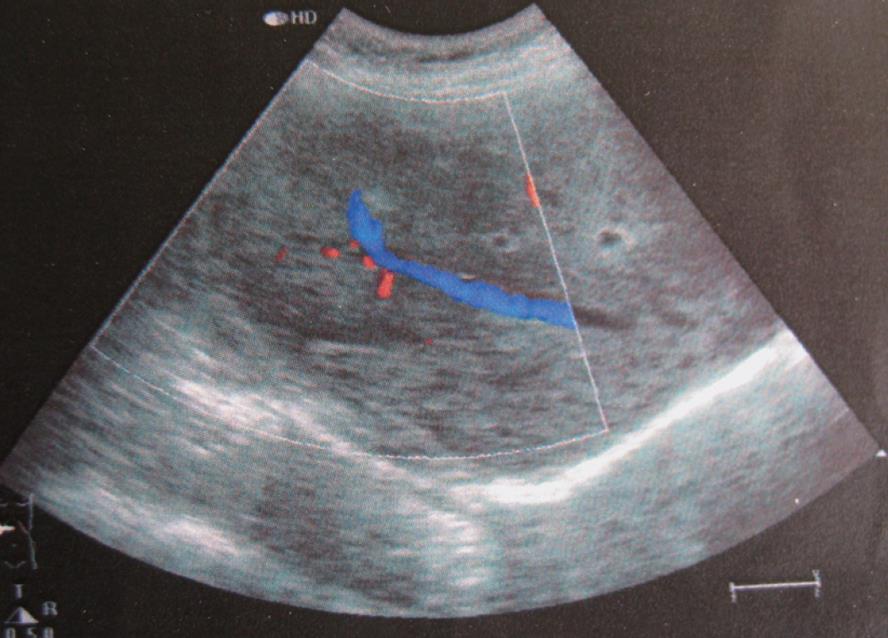
Figure 1 Doppler sonographic image showing a slightly heterogeneous hyperechoic lesion in the right lobe of liver without mass effect on the right hepatic vein.
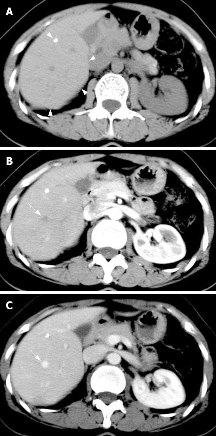
Figure 2 Transverse unenhanced CT image showing the protrudent visceral surface which hints a local iso-attenuated lesion (arrowheads) with punctate calcification (arrow) (A), identical density of the lesion to the adjacent liver parenchyma at arterial phase (B) and portal phase (C).
The right hepatic vein with a normal shape and location crosses the lesion (arrow).
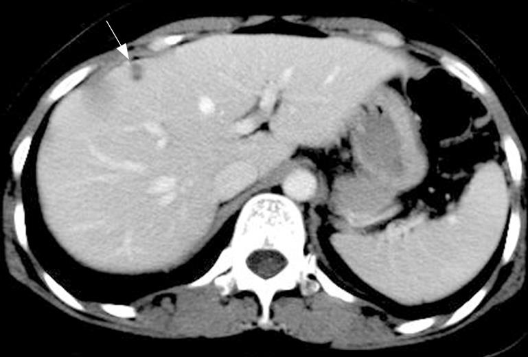
Figure 3 Portal phase image showing a hypo-attenuated lesion (10 mm in diameter) within segment VIII (arrow).
Histopathology proved it to be a gumma.
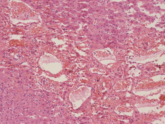
Figure 4 Histology of specimens (HE, × 50).
Microscopy reveals multiple blood-filled cystic spaces and ectatic sinusoids.
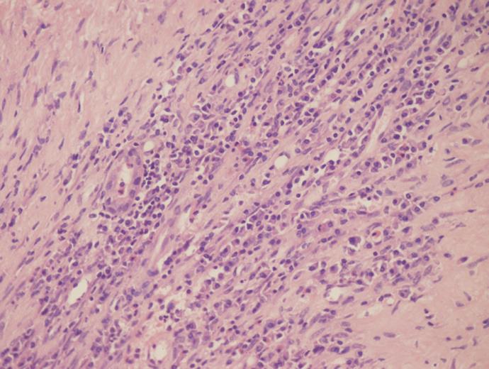
Figure 5 Gumma on segment VIII characterized by multifocal coagulation necrosis and significant infiltration of plasma cells mixed with lymph cells (HE, × 100).
- Citation: Chen JF, Chen WX, Zhang HY, Zhang WY. Peliosis and gummatous syphilis of the liver: A case report. World J Gastroenterol 2008; 14(12): 1961-1963
- URL: https://www.wjgnet.com/1007-9327/full/v14/i12/1961.htm
- DOI: https://dx.doi.org/10.3748/wjg.14.1961


