Copyright
©2008 The WJG Press and Baishideng.
World J Gastroenterol. Mar 14, 2008; 14(10): 1592-1597
Published online Mar 14, 2008. doi: 10.3748/wjg.14.1592
Published online Mar 14, 2008. doi: 10.3748/wjg.14.1592
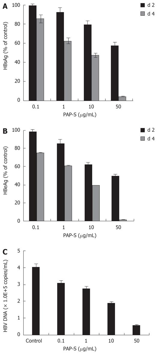
Figure 1 Effects of PAP-S on secreted HBsAg (A), HBeAg (B) and HBV DNA(C) in HepG2 2.
2.15 cell cultures. Data are expressed as mean ± SD of three independent experiments. Except the first group (0.1 &mgr;g/mL), P < 0.01 vs the corresponding controls (Student’s t-test).
Figure 2 Western blot analysis of the expression of pXF3H-PAP in cotransfected HepG2 cells β-actin as internal reference; 1: pHBV1.
3 (1.0 &mgr;g) + pXF3H (2.0 &mgr;g); 2: pHBV1.3 (1.0 &mgr;g) + pXF3H-PAP (2.0 &mgr;g); 3: pHBV1.3 (1.0 &mgr;g) + pXF3H-PAP (1.0 &mgr;g).
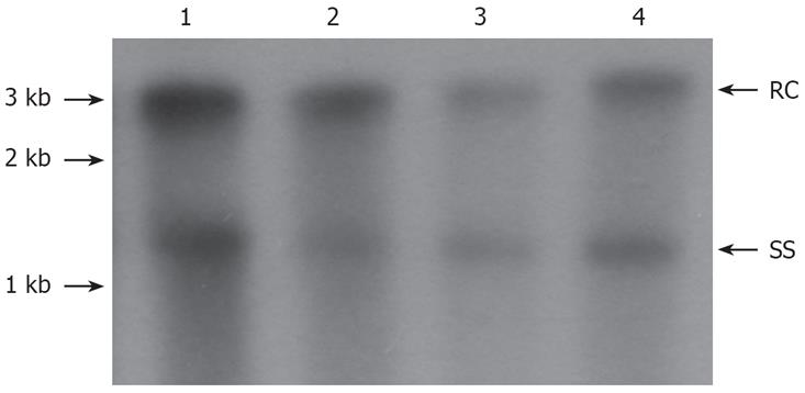
Figure 3 Southern blot analysis of the effect of pXF3H-PAP on intracellular HBV DNA replication in cotransfected HepG2 cells 3 d after transfection.
RC: Relaxed circular DNA; SS: Single stranded DNA. 1: pHBV1.3 (1.0 &mgr;g) + pXF3H (2.0 &mgr;g) 2:pHBV1.3 (1.0 &mgr;g) + pXF3H-PAP (1.0 &mgr;g); 3: pHBV1.3 (1.0 &mgr;g) + pXF3H-PAP (2.0 &mgr;g); 4. pHBV1.3 (1.0 &mgr;g) + 3TC (20.0 &mgr;mol/L).
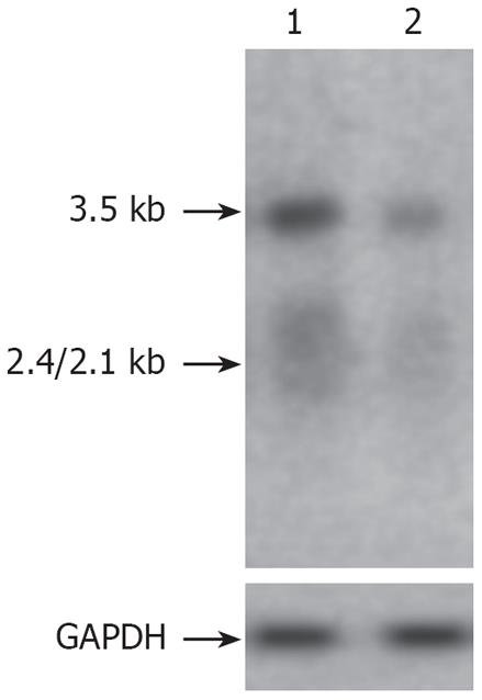
Figure 4 Northern blot analysis of the effect of pXF3H-PAP on intracellular HBV RNA transcription in cotransfected HepG2 cells on d 3 after transfection.
GAPDH as internal reference; 1: pHBV1.3 (1.0 &mgr;g) + pXF3H (2.0 &mgr;g) 2: pHBV1.3 (1.0 &mgr;g) + pXF3H-PAP (2.0 &mgr;g).
-
Citation: He YW, Guo CX, Pan YF, Peng C, Weng ZH. Inhibition of hepatitis B virus replication by pokeweed antiviral protein
in vitro . World J Gastroenterol 2008; 14(10): 1592-1597 - URL: https://www.wjgnet.com/1007-9327/full/v14/i10/1592.htm
- DOI: https://dx.doi.org/10.3748/wjg.14.1592


