Copyright
©2007 Baishideng Publishing Group Co.
World J Gastroenterol. Mar 7, 2007; 13(9): 1372-1377
Published online Mar 7, 2007. doi: 10.3748/wjg.v13.i9.1372
Published online Mar 7, 2007. doi: 10.3748/wjg.v13.i9.1372
Figure 1 A: Typical imported CT image; B: mask applied to highlight stomach.
Figure 2 Schematic diagram indicating how the proximal and distal gastric regions were defined.
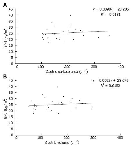
Figure 3 Graphs of regression of BMI on (A) gastric surface area and (B) gastric volume, indicating no linear relationship between these parameters.
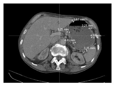
Figure 4 Typical gastric wall thickness measurements on transverse plane CT scan using electronic callipers.
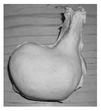
Figure 5 Anatomically correct hollow distensible silicone stomach after wax removal.
-
Citation: Henry JA, O’Sullivan G, Pandit AS. Using computed tomography scans to develop an
ex-vivo gastric model. World J Gastroenterol 2007; 13(9): 1372-1377 - URL: https://www.wjgnet.com/1007-9327/full/v13/i9/1372.htm
- DOI: https://dx.doi.org/10.3748/wjg.v13.i9.1372


