Copyright
©2007 Baishideng Publishing Group Co.
World J Gastroenterol. Feb 14, 2007; 13(6): 882-888
Published online Feb 14, 2007. doi: 10.3748/wjg.v13.i6.882
Published online Feb 14, 2007. doi: 10.3748/wjg.v13.i6.882
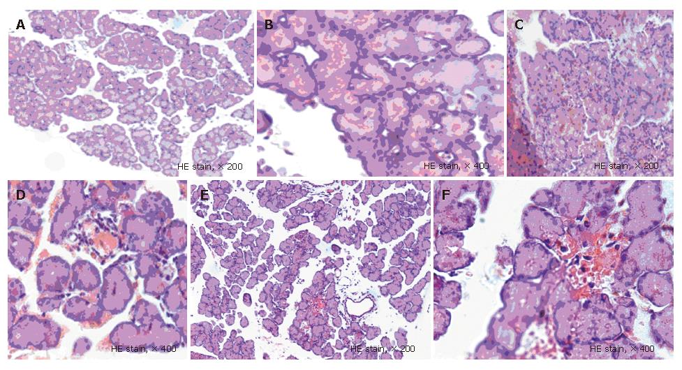
Figure 1 Pathological change of pancreatic tissues at 6 h after operation in each group.
A and B: In NC group, the pancreatic structure was almost normal; C and D: In SAP group, inflammatory cells infiltrated, diffuse bleeding and piecemeal necrosis were occurred; E and F: In BN group, the pathological changes were less serious than those in SAP group.
Figure 2 Expression of NF-κBp65 mRNA at all time points in each group after operation.
A: NC group; B: SAP group; C: BN group. 1: Marker, 2: 1 h, 3: 2 h, 4: 3 h, 5: 6 h, 6: 12 h, 7: 24 h.
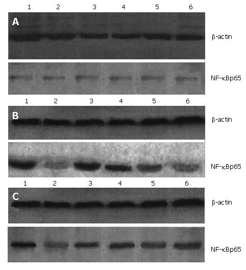
Figure 3 Expression of NF-κBp65 protein at all time points in each group after operation.
A: NC group; B: SAP group; C: BN group. 1: 1 h, 2: 2 h, 3: 3 h, 4: 6 h, 5: 12 h, 6: 24 h.
- Citation: Xia SH, Fang DC, Hu CX, Bi HY, Yang YZ, Di Y. Effect of BN52021 on NFκ-Bp65 expression in pancreatic tissues of rats with severe acute pancreatitis. World J Gastroenterol 2007; 13(6): 882-888
- URL: https://www.wjgnet.com/1007-9327/full/v13/i6/882.htm
- DOI: https://dx.doi.org/10.3748/wjg.v13.i6.882


