Copyright
©2007 Baishideng Publishing Group Inc.
World J Gastroenterol. Nov 7, 2007; 13(41): 5512-5515
Published online Nov 7, 2007. doi: 10.3748/wjg.v13.i41.5512
Published online Nov 7, 2007. doi: 10.3748/wjg.v13.i41.5512
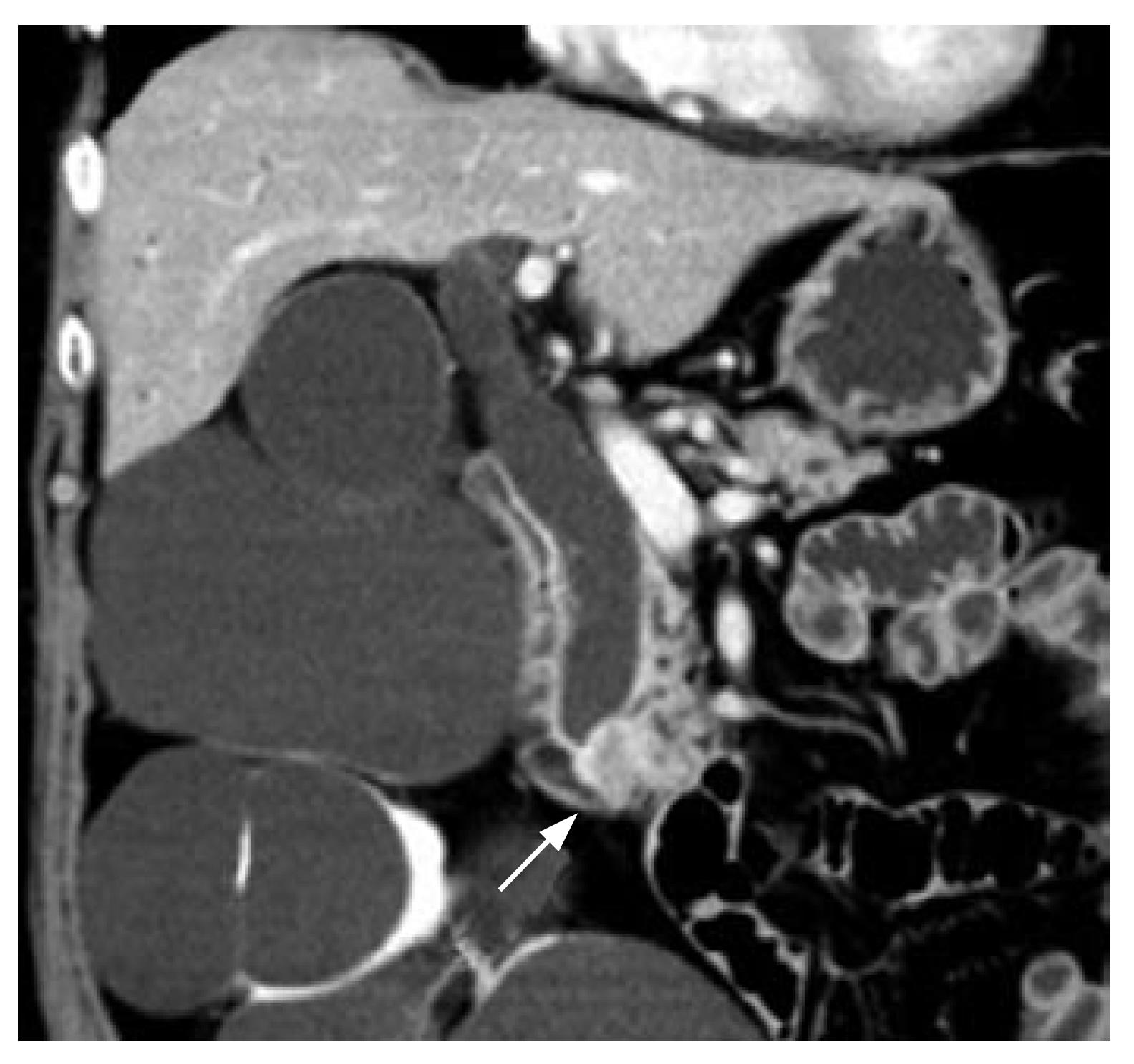
Figure 1 CT reveals a mass in the ampullary region (arrow) with a dilated extrahepatic bile duct.
The patient also had multiple renal cysts.
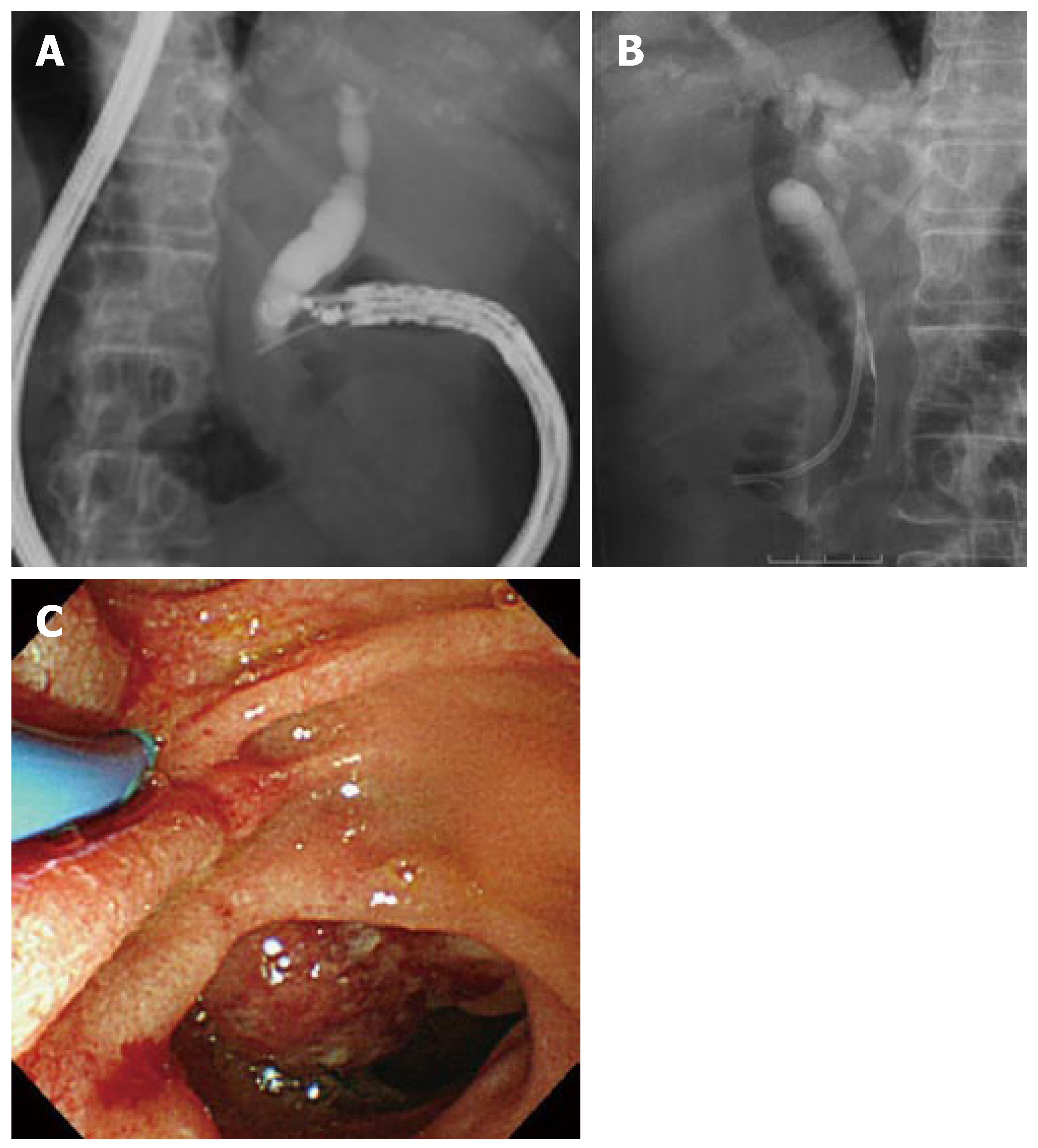
Figure 2 A, B: Puncture of the bile duct via the duodenum under endosonographic guidance, followed by deployment of a plastic stent.
C: Endoscopic view after stent placement.
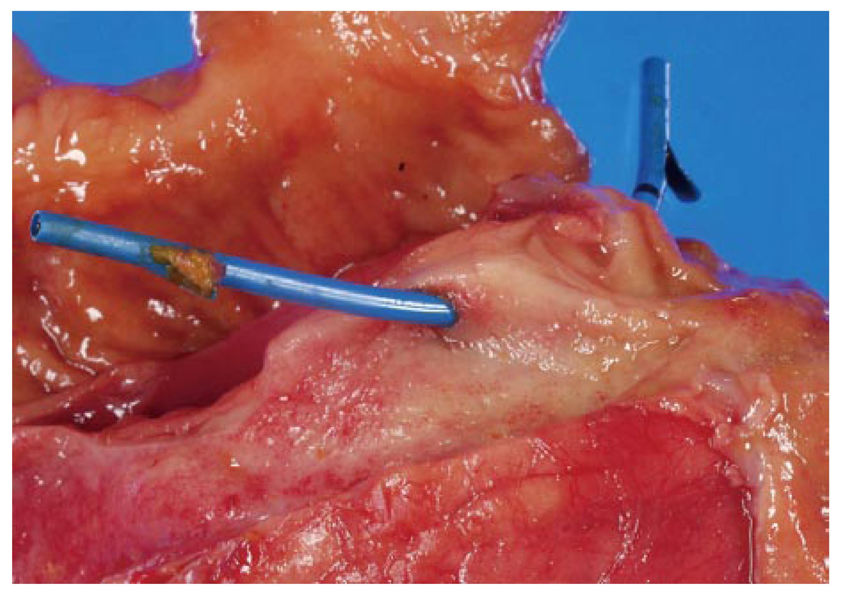
Figure 3 Fresh resected specimen.
No hematoma or abscess is seen at the site of the puncture in the bile duct.
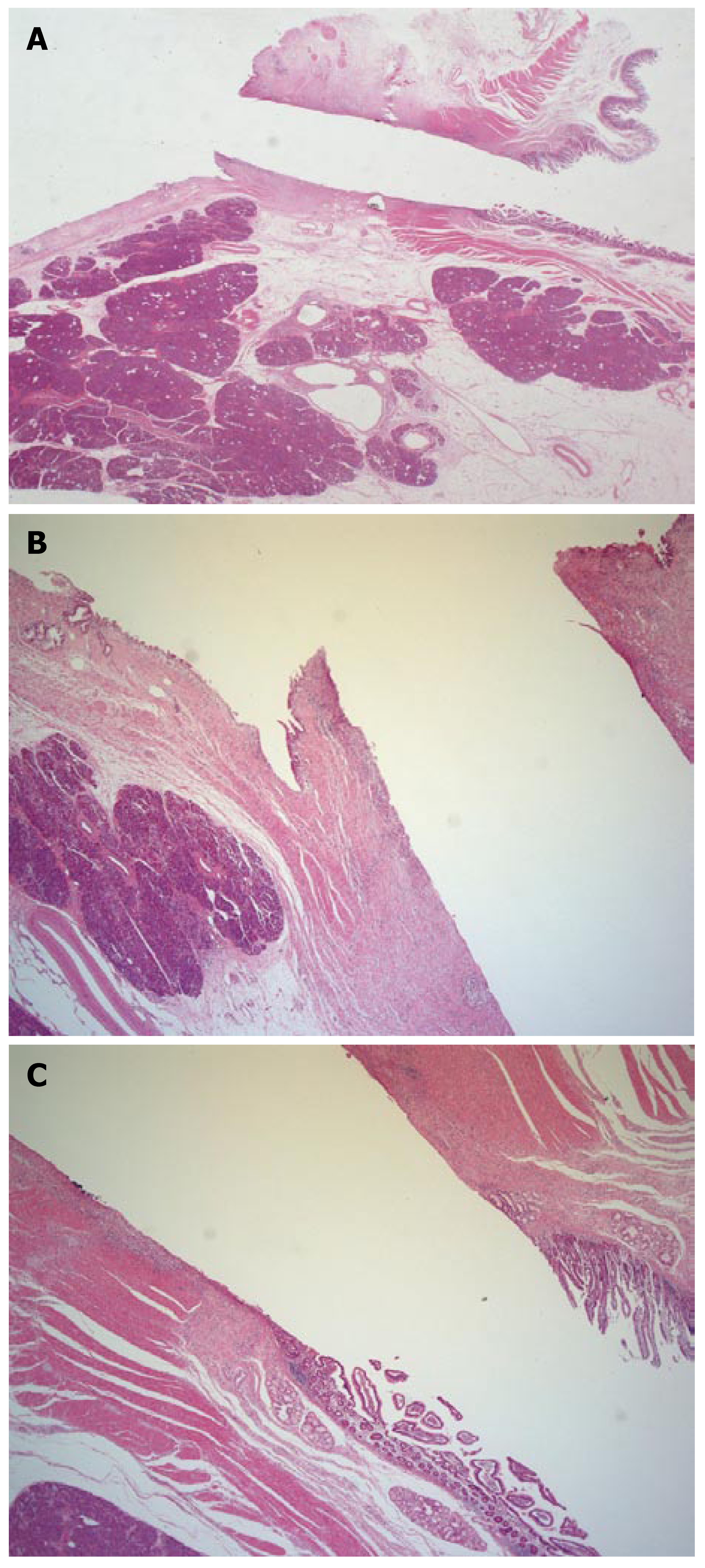
Figure 4 Microscopic views of the sinus tract.
Mild inflammatory cell infiltrate adjacent to the sinus tract in the duodenal wall and the bile duct wall is seen, without hemorrhage or abscess formation. A fistula is formed along the tract of the puncture but without significant reactive changes. A: Low-power view of the sinus tract (× 1.25); B: End of the sinus tract on the bile duct side (× 5); C: End of the sinus tract on the duodenal side (× 5).
- Citation: Fujita N, Noda Y, Kobayashi G, Ito K, Obana T, Horaguchi J, Takasawa O, Nakahara K. Histological changes at an endosonography-guided biliary drainage site: A case report. World J Gastroenterol 2007; 13(41): 5512-5515
- URL: https://www.wjgnet.com/1007-9327/full/v13/i41/5512.htm
- DOI: https://dx.doi.org/10.3748/wjg.v13.i41.5512


