Copyright
©2007 Baishideng Publishing Group Co.
World J Gastroenterol. Jul 28, 2007; 13(28): 3864-3867
Published online Jul 28, 2007. doi: 10.3748/wjg.v13.i28.3864
Published online Jul 28, 2007. doi: 10.3748/wjg.v13.i28.3864
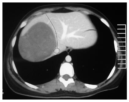
Figure 1 CT image of the hydatic cyst and the hepatic transection line ending with the vena cava inferior at the posterior (black curved line with a ring at the end).
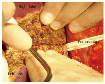
Figure 2 Intraoperative view of the last step of the hepatic transection and the deep transection line (black dashed line).
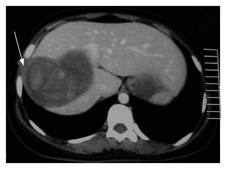
Figure 3 Cystic invasions of both the abdominal wall and the diaphragm (white arrow).
- Citation: Unal A, Pinar Y, Murat Z, Murat K, Ahmet C. A new approach to the surgical treatment of parasitic cysts of the liver: Hepatectomy using the liver hanging maneuver. World J Gastroenterol 2007; 13(28): 3864-3867
- URL: https://www.wjgnet.com/1007-9327/full/v13/i28/3864.htm
- DOI: https://dx.doi.org/10.3748/wjg.v13.i28.3864


