Copyright
©2007 Baishideng Publishing Group Co.
World J Gastroenterol. Jul 28, 2007; 13(28): 3824-3828
Published online Jul 28, 2007. doi: 10.3748/wjg.v13.i28.3824
Published online Jul 28, 2007. doi: 10.3748/wjg.v13.i28.3824
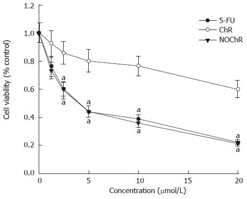
Figure 1 Inhibition of the proliferation of SGC-7901 cells by NOChR.
aP < 0.05 vs treatment with ChR.
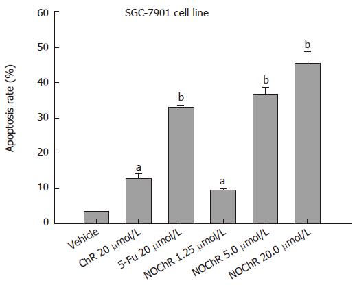
Figure 2 Induction of the apoptosis of SGC-7901 cells by NOChR.
aP < 0.05 vs vehicle; bP < 0.01 vs vehicle.
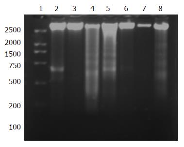
Figure 3 DNA ladder assay showing NOChR-induced apoptosis of SGC-7901 cells.
Lane 1: DNA marker; lane 2: Control; lane 3: 20 μmol/L NOChR (24 h); lane 4: 20 μmol/L NOChR (48 h); lane 5: 20 μmol/L NOChR (72 h); lane 6: 20 μmol/L NOChR plus 10 μmol/L GW9662 (24 h); lane 7: 20 μmol/L NOChR plus 10 μmol/L GW9662 (48 h); lane 8: 20 μmol/L NOChR plus 10 μmol/L GW9662 (72 h).
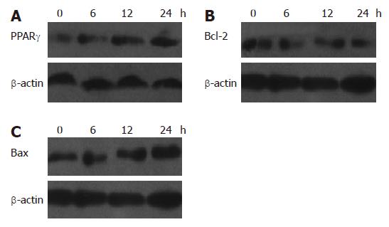
Figure 4 Western blot analysis showing regulation of PPARγ (A), Bcl-2 (B) and Bax (C) protein expressions in SGC-7901 cells by NOChR (mean ± SD, n = 3).
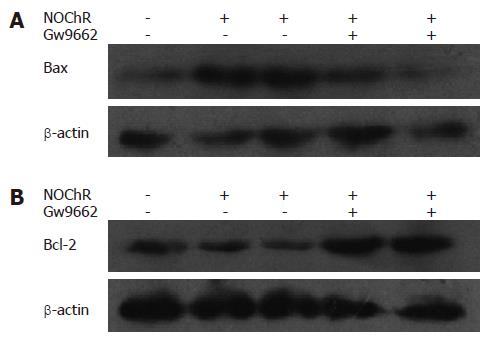
Figure 5 PPARgamma antagonist GW9662 blocked the effects of NOChR on Bax and Bcl-2 protein expression in SGC-7901 cells.
A: Bax; B:Bcl-2. SGC-7901 cells were pretreated with 10 μmol/L GW9662 for 30 min,then exposed to 5, 20 μmol/L NOChR for 24 h respectively (Western blot, mean ± SD, n = 3).
-
Citation: Ai XH, Zheng X, Tang XQ, Sun L, Zhang YQ, Qin Y, Liu HQ, Xia H, Cao JG. Induction of apoptosis of human gastric carcinoma SGC-7901 cell line by 5, 7-dihydroxy-8-nitrochrysin
in vitro . World J Gastroenterol 2007; 13(28): 3824-3828 - URL: https://www.wjgnet.com/1007-9327/full/v13/i28/3824.htm
- DOI: https://dx.doi.org/10.3748/wjg.v13.i28.3824


