Copyright
©2007 Baishideng Publishing Group Co.
World J Gastroenterol. Jul 21, 2007; 13(27): 3773-3775
Published online Jul 21, 2007. doi: 10.3748/wjg.v13.i27.3773
Published online Jul 21, 2007. doi: 10.3748/wjg.v13.i27.3773
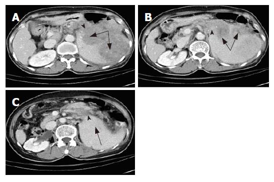
Figure 1 A: Arrow head-pancreatic tail with edematous and swelling changes, with the lesion adhered to the main tumor over the spleen.
Long arrows-hypodense tumor mass occupying the spleen hilum; B: Arrow head-more involvement in the pancreatic tail. Long arrow-the main tumor with mild necrotic change over the spleen; C: Arrow head-the pancreatic tail is still enlarged, with swelling and little fluid acumination. Long arrow-tumor occupying the upper pole of the spleen.
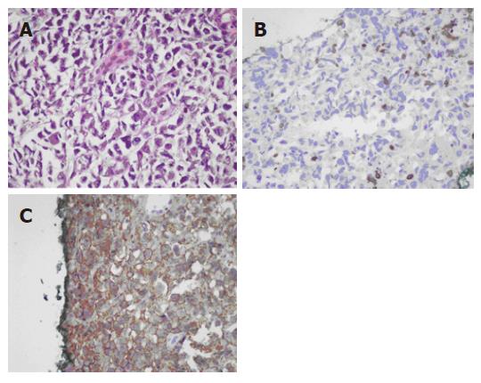
Figure 2 A: Pancreatic tail biopsy showing diffuse large lymphoid cells with highly polymorphic nuclei; B: The neoplastic cells are negative for T cell-associated markers, CD3; C: Large lymphoid cells stained by CD20.
- Citation: Wu CM, Cheng LC, Lo GH, Lai KH, Cheng CL, Pan WC. Malignant lymphoma of spleen presenting as acute pancreatitis: A case report. World J Gastroenterol 2007; 13(27): 3773-3775
- URL: https://www.wjgnet.com/1007-9327/full/v13/i27/3773.htm
- DOI: https://dx.doi.org/10.3748/wjg.v13.i27.3773


